Krahe Daniela D, Woeppel Kevin M, Yang Qianru, Kushwah Neetu, Cui Xinyan Tracy
Department of Bioengineering, University of Pittsburgh, Pittsburgh, PA 15260, USA.
Center for the Neural Basis of Cognition, Pittsburgh, PA 15213, USA.
Antioxidants (Basel). 2022 Aug 22;11(8):1628. doi: 10.3390/antiox11081628.
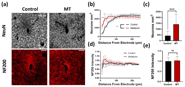
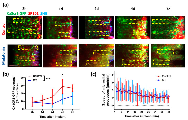
Neural electrode insertion trauma impedes the recording and stimulation capabilities of numerous diagnostic and treatment avenues. Implantation leads to the activation of inflammatory markers and cell types, which is detrimental to neural tissue health and recording capabilities. Oxidative stress and inflammation at the implant site have been shown to decrease with chronic administration of antioxidant melatonin at week 16, but its effects on the acute landscape have not been studied. To assess the effect of melatonin administration in the acute phase, specifically the first week post-implantation, we utilized histological and q-PCR methods to quantify cellular and molecular indicators of inflammation and oxidative stress in the tissue surrounding implanted probes in C57BL/6 mice as well as two-photon microscopy to track the microglial responses to the probes in real-time in transgenic mice expressing GFP with CX3CR1 promotor. Histological results indicate that melatonin effectively maintained neuron density surrounding the electrode, inhibited accumulation and activation of microglia and astrocytes, and reduced oxidative tissue damage. The expression of the pro-inflammatory cytokines, TNF-α and IL-6, were significantly reduced in melatonin-treated animals. Additionally, microglial encapsulation of the implant surface was inhibited by melatonin as compared to control animals following implantation. Our results combined with previous research suggest that melatonin is a particularly suitable drug for modulating inflammatory activity around neural electrode implants both acutely and chronically, translating to more stable and reliable interfaces.
神经电极插入创伤会阻碍多种诊断和治疗途径的记录与刺激能力。植入会导致炎症标志物和细胞类型的激活,这对神经组织健康和记录能力有害。已表明在第16周长期给予抗氧化剂褪黑素后,植入部位的氧化应激和炎症会减轻,但其对急性期情况的影响尚未得到研究。为了评估褪黑素在急性期(特别是植入后的第一周)给药的效果,我们利用组织学和q-PCR方法来量化C57BL/6小鼠植入探针周围组织中炎症和氧化应激的细胞和分子指标,以及利用双光子显微镜实时追踪表达带有CX3CR1启动子的绿色荧光蛋白的转基因小鼠中微胶质细胞对探针的反应。组织学结果表明,褪黑素有效地维持了电极周围的神经元密度,抑制了小胶质细胞和星形胶质细胞的积累和激活,并减少了氧化组织损伤。在褪黑素处理的动物中,促炎细胞因子TNF-α和IL-6的表达显著降低。此外,与植入后的对照动物相比,褪黑素抑制了植入物表面的小胶质细胞包封。我们的结果与先前的研究相结合表明,褪黑素是一种特别适合在急性和慢性期调节神经电极植入物周围炎症活动的药物,这意味着可以实现更稳定和可靠的界面。