Danish Research Centre for Magnetic Resonance, Centre for Functional and Diagnostic Imaging and Research, Copenhagen University Hospital - Amager and Hvidovre, Copenhagen, Denmark.
Danish Research Centre for Magnetic Resonance, Centre for Functional and Diagnostic Imaging and Research, Copenhagen University Hospital - Amager and Hvidovre, Copenhagen, Denmark.
Neuroimage Clin. 2022;36:103147. doi: 10.1016/j.nicl.2022.103147. Epub 2022 Aug 6.
Motor fatigue is common in multiple sclerosis (MS), but its pathophysiology is still poorly understood. Here we used functional magnetic resonance imaging (fMRI) to delineate how the acute induction of motor fatigue alters functional activity of the motor system and how these activity changes are related to motor fatigue.
Forty-four right-handed mildly disabled patients with relapsing-remitting MS and 25 healthy controls performed a maximal tonic precision grip with their right hand until they developed motor fatigue. Before and after the fatiguing task, participants performed a non-fatiguing tonic grip force task, producing 15-20% of their maximum grip force based on visual feedback. Task related brain activity was mapped with blood-oxygen level dependent fMRI at 3 T. Statistical parametric mapping was used to identify relative changes in task-related activation from the pre-fatigue to the recovery MRI session.
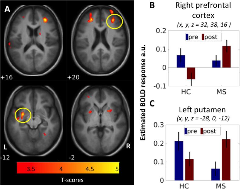
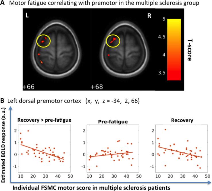
Following fatigue induction, task performance was perturbed in both groups, and task-related activation increased in the right (ipsilateral) primary motor hand area. In patients with MS, task-related activity increased bilaterally during the recovery phase in the ventrolateral portion of the middle putamen and lateral prefrontal cortex relative to controls. The more patients increased task-related activity in left dorsal premotor cortex after the fatiguing task, the less they experienced motor fatigue during daily life.
Patients with MS show enhanced functional engagement of the associative cortico-basal ganglia loop following acute induction of motor fatigue in the contralateral hand. This may reflect increased mental effort to generate movements in the recovery phase after fatigue induction. The ability to recruit the contralateral dorsal premotor cortex after fatigue induction may constitute a protective mechanism against experiencing motor fatigue in everyday life.
运动疲劳在多发性硬化症(MS)中很常见,但它的病理生理学仍知之甚少。在这里,我们使用功能磁共振成像(fMRI)来描绘急性诱导运动疲劳如何改变运动系统的功能活动,以及这些活动变化如何与运动疲劳相关。
44 名右手轻度残疾的复发性缓解型多发性硬化症患者和 25 名健康对照者使用右手进行最大强直精确握力,直到出现运动疲劳。在疲劳任务之前和之后,参与者进行非疲劳强直握力任务,根据视觉反馈产生 15-20%的最大握力。在 3T 磁共振成像上使用血氧水平依赖 fMRI 来绘制任务相关的脑活动。统计参数映射用于从疲劳前 MRI 会话到恢复 MRI 会话识别任务相关激活的相对变化。
疲劳诱导后,两组的任务表现都受到干扰,任务相关的激活在右侧(对侧)初级运动手区增加。在多发性硬化症患者中,与对照组相比,在恢复阶段,双侧中苍白球腹侧和外侧前额叶皮质的任务相关活动增加。在疲劳任务后,患者左背侧运动前皮质的任务相关活动增加越多,他们在日常生活中经历的运动疲劳就越少。
多发性硬化症患者在对侧手急性诱导运动疲劳后表现出联合皮质-基底节环路的功能参与增强。这可能反映了在疲劳诱导后的恢复阶段产生运动时需要增加心理努力。疲劳诱导后对背侧运动前皮质的募集能力可能构成一种对日常生活中体验运动疲劳的保护机制。