• 文献检索
  • 文档翻译
  • 深度研究
  • 学术资讯
  • Suppr Zotero 插件Zotero 插件
  • 邀请有礼
  • 套餐&价格
  • 历史记录
应用&插件
Suppr Zotero 插件Zotero 插件浏览器插件Mac 客户端Windows 客户端微信小程序
定价
高级版会员购买积分包购买API积分包
服务
文献检索文档翻译深度研究API 文档MCP 服务
关于我们
关于 Suppr公司介绍联系我们用户协议隐私条款
关注我们

Suppr 超能文献

核心技术专利:CN118964589B侵权必究
粤ICP备2023148730 号-1Suppr @ 2026

文献检索

告别复杂PubMed语法,用中文像聊天一样搜索,搜遍4000万医学文献。AI智能推荐,让科研检索更轻松。

立即免费搜索

文件翻译

保留排版,准确专业,支持PDF/Word/PPT等文件格式,支持 12+语言互译。

免费翻译文档

深度研究

AI帮你快速写综述,25分钟生成高质量综述,智能提取关键信息,辅助科研写作。

立即免费体验

慢性胰腺炎的定量 MRI:多机构前瞻性研究结果,磁共振成像作为评估胰腺纤维化的非侵入性方法 (MINIMAP)。

Quantitative MRI of chronic pancreatitis: results from a multi-institutional prospective study, magnetic resonance imaging as a non-invasive method for assessment of pancreatic fibrosis (MINIMAP).

机构信息

Department of Radiology and Imaging Sciences, Indiana University School of Medicine Indianapolis, 550 N. University Blvd. Suite 0663, Indianapolis, IN, 46202, USA.

Department of Medicine Division of Gastroenterology, Hepatology & Nutrition University of Pittsburgh School of Medicine, Pittsburgh, PA, USA.

出版信息

Abdom Radiol (NY). 2022 Nov;47(11):3792-3805. doi: 10.1007/s00261-022-03654-7. Epub 2022 Aug 29.

DOI:
10.1007/s00261-022-03654-7
PMID:36038644
原文链接:https://pmc.ncbi.nlm.nih.gov/articles/PMC9423890/
Abstract

PURPOSE

To determine if quantitative MRI techniques can be helpful to evaluate chronic pancreatitis (CP) in a setting of multi-institutional study.

METHODS

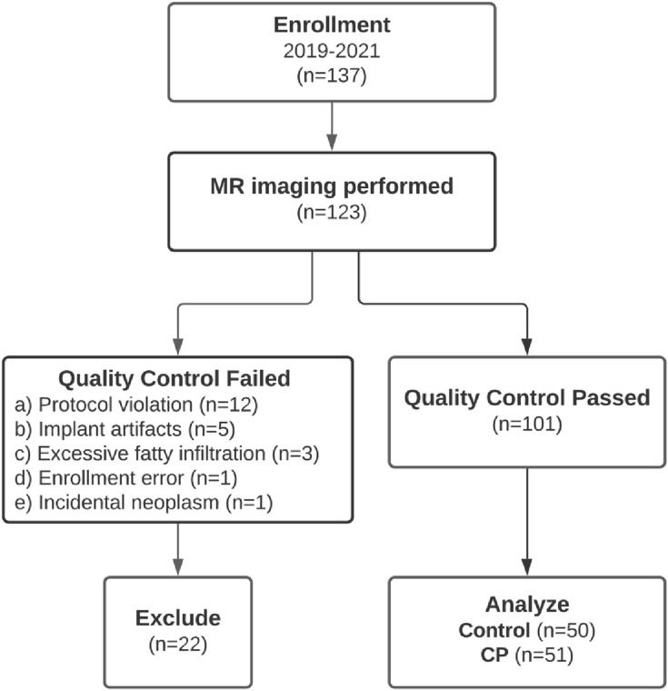
This study included a subgroup of participants (n = 101) enrolled in the Prospective Evaluation of Chronic Pancreatitis for Epidemiologic and Translational Studies (PROCEED) study (NCT03099850) from February 2019 to May 2021. MRI was performed on 1.5 T using Siemens and GE scanners at seven clinical centers across the USA. Quantitative MRI parameters of the pancreas included T1 relaxation time, extracellular volume (ECV) fraction, apparent diffusion coefficient (ADC), and fat signal fraction. We report the diagnostic performance and mean values within the control (n = 50) and CP (n = 51) groups. The T1, ECV and fat signal fraction were combined to generate the quantitative MRI score (Q-MRI).

RESULTS

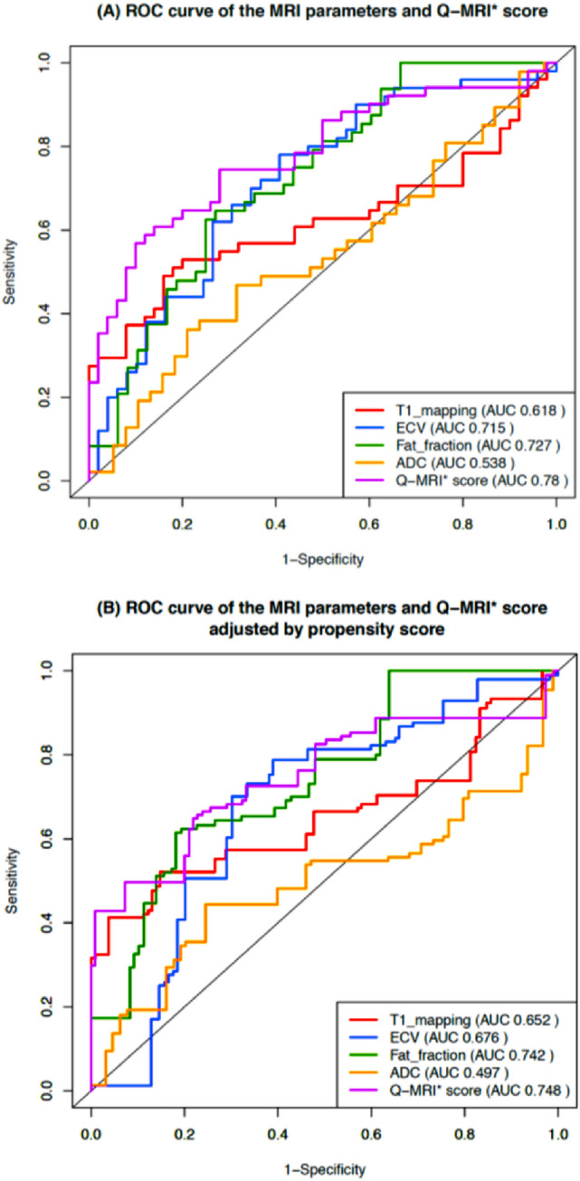
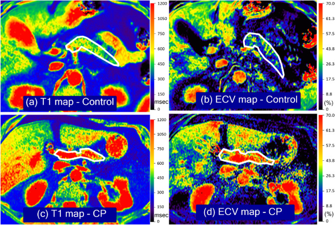
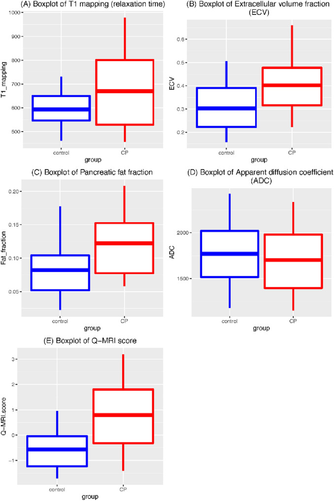
There was significantly higher T1 relaxation time; mean 669 ms (± 171) vs. 593 ms (± 82) (p = 0.006), ECV fraction; 40.2% (± 14.7) vs. 30.3% (± 11.9) (p < 0.001), and pancreatic fat signal fraction; 12.2% (± 5.5) vs. 8.2% (± 4.4) (p < 0.001) in the CP group compared to controls. The ADC was similar between groups (p = 0.45). The AUCs for the T1, ECV, and pancreatic fat signal fraction were 0.62, 0.72, and 0.73, respectively. The composite Q-MRI score improved the diagnostic performance (cross-validated AUC: 0.76).

CONCLUSION

Quantitative MR parameters evaluating the pancreatic parenchyma (T1, ECV fraction, and fat signal fraction) are helpful in the diagnosis of CP. A Q-MRI score that combines these three MR parameters improves diagnostic performance. Further studies are warranted with larger study populations including patients with acute and recurrent acute pancreatitis and longitudinal follow-ups.

摘要

目的

确定定量 MRI 技术是否有助于在多机构研究中评估慢性胰腺炎(CP)。

方法

本研究纳入了 2019 年 2 月至 2021 年 5 月期间参与 Prospective Evaluation of Chronic Pancreatitis for Epidemiologic and Translational Studies(PROCEED)研究(NCT03099850)的参与者亚组(n=101)。该研究在 7 个美国临床中心的西门子和 GE 扫描仪上进行了 1.5T MRI 检查。胰腺的定量 MRI 参数包括 T1 弛豫时间、细胞外体积(ECV)分数、表观扩散系数(ADC)和脂肪信号分数。我们报告了对照组(n=50)和 CP 组(n=51)的诊断性能和平均值。T1、ECV 和脂肪信号分数结合起来生成定量 MRI 评分(Q-MRI)。

结果

CP 组的 T1 弛豫时间显著升高,平均为 669ms(±171)vs. 593ms(±82)(p=0.006),ECV 分数为 40.2%(±14.7)vs. 30.3%(±11.9)(p<0.001),胰腺脂肪信号分数为 12.2%(±5.5)vs. 8.2%(±4.4)(p<0.001)。与对照组相比,CP 组的 ADC 相似(p=0.45)。T1、ECV 和胰腺脂肪信号分数的 AUC 分别为 0.62、0.72 和 0.73。复合 Q-MRI 评分提高了诊断性能(交叉验证 AUC:0.76)。

结论

评估胰腺实质的定量 MRI 参数(T1、ECV 分数和脂肪信号分数)有助于 CP 的诊断。结合这三个 MRI 参数的 Q-MRI 评分可提高诊断性能。需要进一步研究,纳入更多的急性和复发性胰腺炎患者以及进行纵向随访。

https://cdn.ncbi.nlm.nih.gov/pmc/blobs/5d69/9423890/96d17516c1a0/261_2022_3654_Fig4_HTML.jpg
https://cdn.ncbi.nlm.nih.gov/pmc/blobs/5d69/9423890/ce770c7cf2d1/261_2022_3654_Fig1_HTML.jpg
https://cdn.ncbi.nlm.nih.gov/pmc/blobs/5d69/9423890/0e378bbba35e/261_2022_3654_Fig2_HTML.jpg
https://cdn.ncbi.nlm.nih.gov/pmc/blobs/5d69/9423890/94f7f3806c54/261_2022_3654_Fig3_HTML.jpg
https://cdn.ncbi.nlm.nih.gov/pmc/blobs/5d69/9423890/96d17516c1a0/261_2022_3654_Fig4_HTML.jpg

相似文献

1
Quantitative MRI of chronic pancreatitis: results from a multi-institutional prospective study, magnetic resonance imaging as a non-invasive method for assessment of pancreatic fibrosis (MINIMAP).慢性胰腺炎的定量 MRI:多机构前瞻性研究结果,磁共振成像作为评估胰腺纤维化的非侵入性方法 (MINIMAP)。
Abdom Radiol (NY). 2022 Nov;47(11):3792-3805. doi: 10.1007/s00261-022-03654-7. Epub 2022 Aug 29.
2
Diagnosis of chronic pancreatitis using semi-quantitative MRI features of the pancreatic parenchyma: results from the multi-institutional MINIMAP study.使用胰腺实质的半定量 MRI 特征诊断慢性胰腺炎:来自多机构 MINIMAP 研究的结果。
Abdom Radiol (NY). 2023 Oct;48(10):3162-3173. doi: 10.1007/s00261-023-04000-1. Epub 2023 Jul 12.
3
Magnetic resonance imaging as a non-invasive method for the assessment of pancreatic fibrosis (MINIMAP): a comprehensive study design from the consortium for the study of chronic pancreatitis, diabetes, and pancreatic cancer.磁共振成像作为一种评估胰腺纤维化的非侵入性方法 (MINIMAP):来自慢性胰腺炎、糖尿病和胰腺癌研究联盟的综合研究设计。
Abdom Radiol (NY). 2019 Aug;44(8):2809-2821. doi: 10.1007/s00261-019-02049-5.
4
T1 relaxation times and MR elastography-derived stiffness: new potential imaging biomarkers for the assessment of chronic pancreatitis.T1 弛豫时间和 MR 弹性成像衍生的刚性:评估慢性胰腺炎的新潜在成像生物标志物。
Abdom Radiol (NY). 2021 Dec;46(12):5598-5608. doi: 10.1007/s00261-021-03276-5. Epub 2021 Sep 15.
5
Single- and multiparameter magnetic resonance imaging for diagnosing and severity grading of chronic pancreatitis.用于慢性胰腺炎诊断及严重程度分级的单参数和多参数磁共振成像
Abdom Radiol (NY). 2023 Feb;48(2):630-641. doi: 10.1007/s00261-022-03760-6. Epub 2022 Dec 7.
6
Progression of parenchymal and ductal findings in patients with chronic pancreatitis: A 4-year follow-up MRI study.慢性胰腺炎患者实质和导管病变的进展:一项为期 4 年的 MRI 随访研究。
Eur J Radiol. 2020 Apr;125:108868. doi: 10.1016/j.ejrad.2020.108868. Epub 2020 Feb 7.
7
Quantitative MRI of the liver: Evaluation of extracellular volume fraction and other quantitative parameters in comparison to MR elastography for the assessment of hepatopathy.肝脏的定量磁共振成像:对比磁共振弹性成像评估肝纤维化的细胞外容积分数和其他定量参数
Magn Reson Imaging. 2021 Apr;77:7-13. doi: 10.1016/j.mri.2020.12.005. Epub 2020 Dec 9.
8
Secretin-stimulated MRI characterization of pancreatic morphology and function in patients with chronic pancreatitis.慢性胰腺炎患者中促胰液素刺激下胰腺形态和功能的磁共振成像特征
Pancreatology. 2017 Mar-Apr;17(2):228-236. doi: 10.1016/j.pan.2017.01.009. Epub 2017 Jan 27.
9
Multiparametric MRI Scoring System of the Pancreas for the Diagnosis of Chronic Pancreatitis.用于诊断慢性胰腺炎的胰腺多参数磁共振成像评分系统
J Magn Reson Imaging. 2025 May;61(5):2183-2194. doi: 10.1002/jmri.29594. Epub 2024 Sep 3.
10
Quantitative MR Evaluation of Chronic Pancreatitis: Extracellular Volume Fraction and MR Relaxometry.慢性胰腺炎的定量磁共振评估:细胞外容积分数和磁共振弛豫率。
AJR Am J Roentgenol. 2018 Mar;210(3):533-542. doi: 10.2214/AJR.17.18606. Epub 2018 Jan 16.

引用本文的文献

1
Obesity and Pancreatic Diseases: From Inflammation to Oncogenesis and the Impact of Weight Loss Interventions.肥胖与胰腺疾病:从炎症到肿瘤发生以及减肥干预措施的影响
Nutrients. 2025 Jul 14;17(14):2310. doi: 10.3390/nu17142310.
2
Automated pancreatic extracellular volume fraction as a biomarker for microangiopathy in type 2 diabetes mellitus.自动化胰腺细胞外容积分数作为2型糖尿病微血管病变的生物标志物
BMC Med Imaging. 2025 Jul 1;25(1):226. doi: 10.1186/s12880-025-01731-z.
3
Whole pancreas water T mapping at 3 Tesla.3特斯拉下的全胰腺水T成像

本文引用的文献

1
Simulated Reduced-Count Whole-Body FDG PET: Evaluation in Children and Young Adults Imaged on a Digital PET Scanner.模拟低计数全身 FDG PET:数字化 PET 扫描仪在儿童和青年受检者中的评估。
AJR Am J Roentgenol. 2022 Dec;219(6):952-961. doi: 10.2214/AJR.22.27894. Epub 2022 Jun 22.
2
Evaluation of Pancreatic Fibrosis Grading by Multiparametric Quantitative Magnetic Resonance Imaging.多参数定量磁共振成像评估胰腺纤维化分级。
J Magn Reson Imaging. 2021 Nov;54(5):1417-1429. doi: 10.1002/jmri.27626. Epub 2021 Apr 5.
3
Image-Based Machine Learning Algorithms for Disease Characterization in the Human Type 1 Diabetes Pancreas.
MAGMA. 2025 Apr;38(2):271-283. doi: 10.1007/s10334-025-01224-8. Epub 2025 Mar 6.
4
Association between intra-pancreatic fat deposition and diseases of the exocrine pancreas: A narrative review.胰腺内脂肪沉积与胰腺外分泌疾病之间的关联:一项叙述性综述。
World J Gastroenterol. 2025 Jan 14;31(2):101180. doi: 10.3748/wjg.v31.i2.101180.
5
Pancreatic volume, fat content and T1 relaxation time using magnetic resonance imaging in patients with prior gestational diabetes mellitus.使用磁共振成像对既往有妊娠糖尿病患者的胰腺体积、脂肪含量及T1弛豫时间进行研究。
Abdom Radiol (NY). 2025 Jun;50(6):2649-2659. doi: 10.1007/s00261-024-04698-7. Epub 2024 Nov 27.
6
Multiparametric MRI Scoring System of the Pancreas for the Diagnosis of Chronic Pancreatitis.用于诊断慢性胰腺炎的胰腺多参数磁共振成像评分系统
J Magn Reson Imaging. 2025 May;61(5):2183-2194. doi: 10.1002/jmri.29594. Epub 2024 Sep 3.
7
Advances in MRI of Chronic Pancreatitis.慢性胰腺炎的磁共振成像进展
Adv Clin Radiol. 2024 Sep;6(1):31-39. doi: 10.1016/j.yacr.2024.04.002. Epub 2024 Jun 15.
8
3D histology reveals that immune response to pancreatic precancers is heterogeneous and depends on global pancreas structure.三维组织学显示,对胰腺癌前病变的免疫反应是异质性的,并且取决于胰腺的整体结构。
bioRxiv. 2024 Aug 6:2024.08.03.606493. doi: 10.1101/2024.08.03.606493.
9
Imaging abnormalities of the pancreas in diabetes: implications for diagnosis and treatment.糖尿病患者胰腺的影像学异常:对诊断和治疗的影响。
Curr Opin Gastroenterol. 2024 Sep 1;40(5):381-388. doi: 10.1097/MOG.0000000000001054. Epub 2024 Jun 28.
10
T1 and T2-mapping in pancreatic MRI: Current evidence and future perspectives.胰腺MRI中的T1和T2映射:当前证据与未来展望。
Eur J Radiol Open. 2024 May 30;12:100572. doi: 10.1016/j.ejro.2024.100572. eCollection 2024 Jun.
基于图像的机器学习算法在人类 1 型糖尿病胰腺疾病特征中的应用。
Am J Pathol. 2021 Mar;191(3):454-462. doi: 10.1016/j.ajpath.2020.11.010. Epub 2020 Dec 8.
4
Quantitative assessment of disease severity of primary sclerosing cholangitis with T1 mapping and extracellular volume imaging.利用T1映射和细胞外容积成像对原发性硬化性胆管炎疾病严重程度进行定量评估。
Abdom Radiol (NY). 2021 Jun;46(6):2433-2443. doi: 10.1007/s00261-020-02839-2. Epub 2020 Nov 1.
5
Organisation of the human pancreas in health and in diabetes.人胰腺在健康和糖尿病中的组织。
Diabetologia. 2020 Oct;63(10):1966-1973. doi: 10.1007/s00125-020-05203-7. Epub 2020 Sep 7.
6
Is Cambridge scoring in chronic pancreatitis the same using ERCP and MRCP?: A need for revision of standards.剑桥评分系统在 ERCP 和 MRCP 中用于慢性胰腺炎的诊断是否相同?:需要修订标准。
Abdom Radiol (NY). 2021 Feb;46(2):647-654. doi: 10.1007/s00261-020-02685-2. Epub 2020 Aug 1.
7
Inter-observer variability of radiologists for Cambridge classification of chronic pancreatitis using CT and MRCP: results from a large multi-center study.放射科医生使用 CT 和 MRCP 对慢性胰腺炎剑桥分类的观察者间变异性:一项大型多中心研究的结果。
Abdom Radiol (NY). 2020 May;45(5):1481-1487. doi: 10.1007/s00261-020-02521-7.
8
Progression of parenchymal and ductal findings in patients with chronic pancreatitis: A 4-year follow-up MRI study.慢性胰腺炎患者实质和导管病变的进展:一项为期 4 年的 MRI 随访研究。
Eur J Radiol. 2020 Apr;125:108868. doi: 10.1016/j.ejrad.2020.108868. Epub 2020 Feb 7.
9
ACG Clinical Guideline: Chronic Pancreatitis.ACG 临床指南:慢性胰腺炎。
Am J Gastroenterol. 2020 Mar;115(3):322-339. doi: 10.14309/ajg.0000000000000535.
10
Evaluation of variable flip angle, MOLLI, SASHA, and IR-SNAPSHOT pulse sequences for T relaxometry and extracellular volume imaging of the pancreas and liver.评估可变翻转角、MOLLI、SASHA 和 IR-SNAPSHOT 脉冲序列在胰腺和肝脏 T 弛豫测量和细胞外容积成像中的应用。
MAGMA. 2019 Oct;32(5):559-566. doi: 10.1007/s10334-019-00762-2. Epub 2019 Jun 4.