Department of Morphology, School of Medicine, Federal University of Ceará, Fortaleza, Ceará, Brazil.
Paulo Niemeyer Brain Institute, Federal University of Rio de Janeiro, UFRJ, Rio de Janeiro, Rio de Janeiro, Brazil.
Front Immunol. 2022 Aug 22;13:956340. doi: 10.3389/fimmu.2022.956340. eCollection 2022.
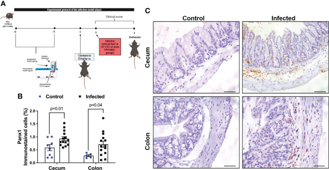
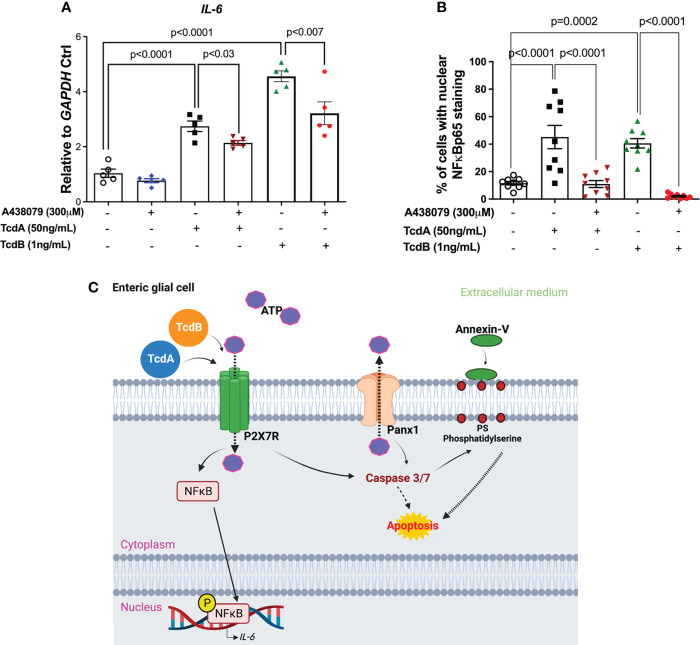
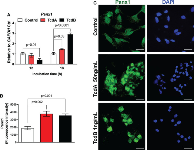
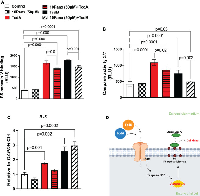
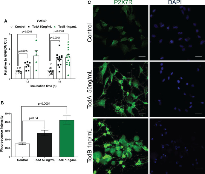
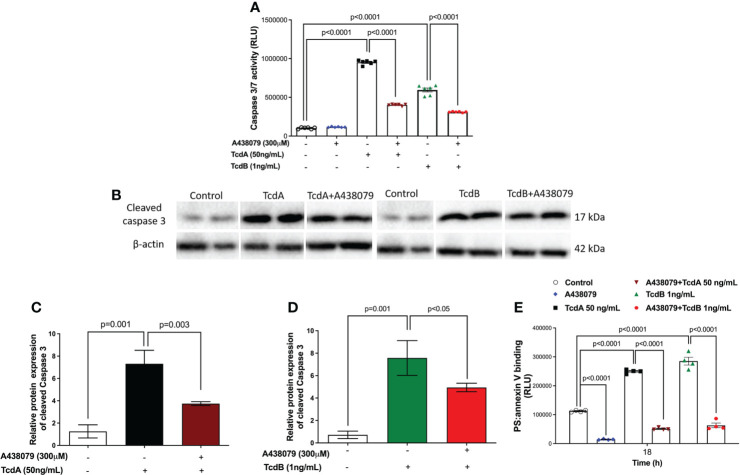
( produces toxins A (TcdA) and B (TcdB), both associated with intestinal damage and diarrhea. Pannexin-1 (Panx1) channels allows the passage of messenger molecules, such as adenosine triphosphate (ATP), which in turn activate the P2X7 receptors (P2X7R) that regulate inflammation and cell death in inflammatory bowel diseases. The aim of this study was to verify the effect of infection (CDI) in the expression of Panx1 and P2X7R in intestinal tissues of mice, as well as their role in cell death and expression induced by TcdA and TcdB in enteric glial cells (EGCs). Male C57BL/6 mice (8 weeks of age) were infected with VPI10463, and the control group received only vehicle per gavage. After three days post-infection (p.i.), cecum and colon samples were collected to evaluate the expression of Panx1 by immunohistochemistry. , EGCs (PK060399egfr) were challenged with TcdA or TcdB, in the presence or absence of the Panx1 inhibitor (10Panx trifluoroacetate) or P2X7R antagonist (A438079), and Panx1 and P2X7R expression, caspase-3/7 activity and phosphatidylserine binding to annexin-V, as well as expression were assessed. CDI increased the levels of Panx1 in cecum and colon of mice compared to the control group. Panx1 inhibitor decreased caspase-3/7 activity and phosphatidylserine-annexin-V binding, but not gene expression in TcdA and TcdB-challenged EGCs. P2X7 receptor antagonist accentually reduced caspase-3/7 activity, phosphatidylserine-annexin-V binding, and gene expression in TcdA and TcdB-challenged EGCs. In conclusion, Panx1 is increased during CDI and plays an important role in the effects of toxins in EGCs, participating in cell death induced by both toxins by promoting caspase-3/7 activation P2X7R, which is also involved in IL-6 expression induced by both toxins.
(产生毒素 A(TcdA)和 B(TcdB),两者均与肠道损伤和腹泻有关。连接蛋白 1(Panx1)通道允许信使分子如三磷酸腺苷(ATP)通过,后者反过来激活调节炎症性肠病中炎症和细胞死亡的 P2X7 受体(P2X7R)。本研究旨在验证 感染(CDI)对小鼠肠组织中 Panx1 和 P2X7R 表达的影响,以及它们在 TcdA 和 TcdB 诱导的肠嗜铬细胞(EGCs)死亡和 表达中的作用。雄性 C57BL/6 小鼠(8 周龄)用 VPI10463 感染,对照组仅用灌胃给予载体。感染后 3 天(p.i.),收集盲肠和结肠样本,通过免疫组织化学评估 Panx1 的表达。此外,用 TcdA 或 TcdB 挑战 EGC(PK060399egfr),存在或不存在 Panx1 抑制剂(10Panx 三氟乙酸盐)或 P2X7R 拮抗剂(A438079),并评估 Panx1 和 P2X7R 表达、半胱天冬酶-3/7 活性和磷脂酰丝氨酸与 annexin-V 的结合以及 表达。与对照组相比,CDI 增加了小鼠盲肠和结肠中 Panx1 的水平。Panx1 抑制剂降低了 TcdA 和 TcdB 挑战的 EGC 中 caspase-3/7 活性和磷脂酰丝氨酸-annexin-V 结合,但不降低 基因表达。P2X7 受体拮抗剂明显降低了 TcdA 和 TcdB 挑战的 EGC 中 caspase-3/7 活性、磷脂酰丝氨酸-annexin-V 结合和 基因表达。总之,CDI 期间 Panx1 增加,并在 毒素对 EGC 的作用中发挥重要作用,通过促进 caspase-3/7 激活和 P2X7R 参与两种毒素诱导的细胞死亡,后者也参与两种毒素诱导的 IL-6 表达。