CellSight Technologies Incorporated, San Francisco, California, USA.
Department of Radiology, Stanford University, Palo Alto, California, USA.
Mol Imaging. 2022 Aug 8;2022:3667417. doi: 10.1155/2022/3667417. eCollection 2022.
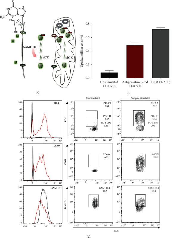
[F]F-AraG is a radiolabeled nucleoside analog that shows relative specificity for activated T cells. The aim of this study was to investigate the biodistribution of [F]F-AraG in healthy volunteers and assess the preliminary safety and radiation dosimetry.
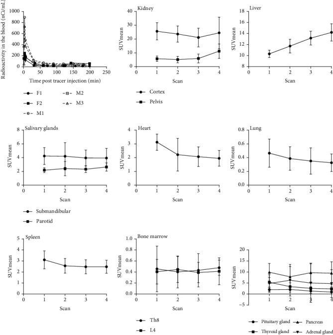
Six healthy subjects (three female and three male) between the ages of 24 and 60 participated in the study. Each subject received a bolus venous injection of [F]F-AraG (dose range: 244.2-329.3 MBq) prior to four consecutive PET/MR whole-body scans. Blood samples were collected at regular intervals and vital signs monitored before and after tracer administration. Regions of interest were delineated for multiple organs, and the area under the time-activity curves was calculated for each organ and used to derive time-integrated activity coefficient (TIAC). TIACs were input for absorbed dose and effective dose calculations using OLINDA.
PET/MR examination was well tolerated, and no adverse effects to the administration of [F]F-AraG were noted by the study participants. The biodistribution was generally reflective of the expression and activity profiles of the enzymes involved in [F]F-AraG's cellular accumulation, mitochondrial kinase dGK, and SAMHD1. The highest uptake was observed in the kidneys and liver, while the brain, lung, bone marrow, and muscle showed low tracer uptake. The estimated effective dose for [F]F-AraG was 0.0162 mSv/MBq (0.0167 mSv/MBq for females and 0.0157 mSv/MBq for males).
Biodistribution of [F]F-AraG in healthy volunteers was consistent with its association with mitochondrial metabolism. PET/MR [F]F-AraG imaging was well tolerated, with a radiation dosimetry profile similar to other commonly used [F]-labeled tracers. [F]F-AraG's connection with mitochondrial biogenesis and favorable biodistribution characteristics make it an attractive tracer with a variety of potential applications.
[F]F-AraG 是一种放射性标记的核苷类似物,对活化的 T 细胞具有相对特异性。本研究旨在研究健康志愿者中[F]F-AraG 的生物分布,并评估初步安全性和辐射剂量学。
6 名年龄在 24 岁至 60 岁之间的健康志愿者(3 名女性和 3 名男性)参与了这项研究。每位志愿者在进行连续 4 次 PET/MR 全身扫描前,静脉注射[F]F-AraG(剂量范围:244.2-329.3MBq)。在给予示踪剂前后定期采集血样并监测生命体征。为多个器官划定感兴趣区,并计算每个器官的时间-活性曲线下面积,用于推导时间积分活度系数(TIAC)。使用 OLINDA 将 TIAC 输入到吸收剂量和有效剂量计算中。
PET/MR 检查耐受良好,研究参与者未观察到[F]F-AraG 给药的不良反应。生物分布总体上反映了参与[F]F-AraG 细胞积累的酶、线粒体激酶 dGK 和 SAMHD1 的表达和活性谱。摄取最高的器官是肾脏和肝脏,而大脑、肺、骨髓和肌肉摄取示踪剂的水平较低。[F]F-AraG 的估计有效剂量为 0.0162mSv/MBq(女性为 0.0167mSv/MBq,男性为 0.0157mSv/MBq)。
健康志愿者中[F]F-AraG 的生物分布与其与线粒体代谢的关联一致。PET/MR [F]F-AraG 成像耐受良好,辐射剂量学特征与其他常用[F]-标记示踪剂相似。[F]F-AraG 与线粒体生物发生的联系以及有利的生物分布特征使其成为一种有吸引力的示踪剂,具有多种潜在应用。