Department of Physical Therapy and Rehabilitation Science, University of California, San Francisco, San Francisco, California;
Department of Radiology and Biomedical Imaging, University of California, San Francisco, San Francisco, California.
J Nucl Med. 2022 Jan;63(1):140-146. doi: 10.2967/jnumed.120.259325. Epub 2021 Apr 9.
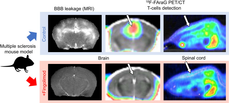
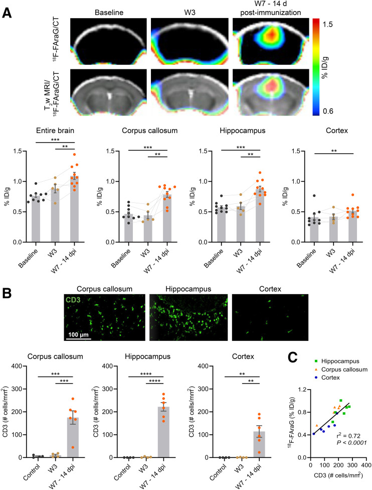
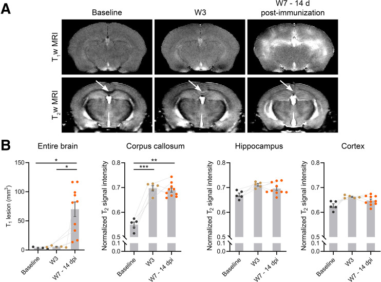
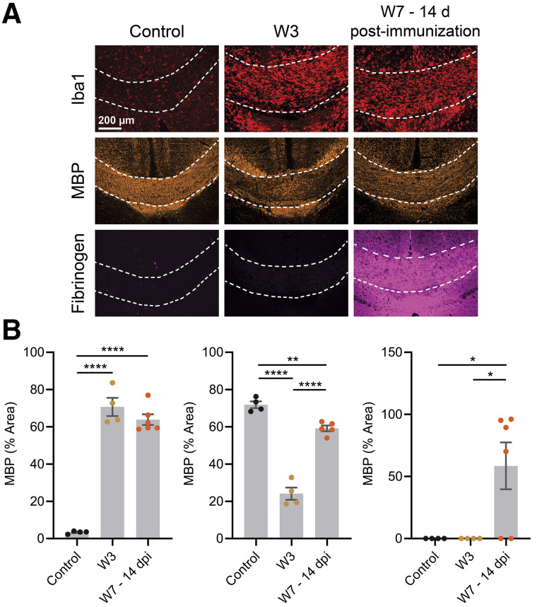
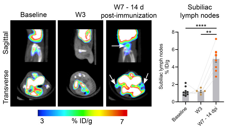
Lymphocytes and innate immune cells are key drivers of multiple sclerosis (MS) and are the main target of MS disease-modifying therapies (DMT). Ex vivo analyses of MS lesions have revealed cellular heterogeneity and variable T cell levels, which may have important implications for patient stratification and choice of DMT. Although MRI has proven valuable to monitor DMT efficacy, its lack of specificity for cellular subtypes highlights the need for complementary methods to improve lesion characterization. Here, we evaluated the potential of 2'-deoxy-2'-F-fluoro-9-β-d-arabinofuranosylguanine (F-FAraG) PET imaging to noninvasively assess infiltrating T cells and to provide, in combination with MRI, a novel tool to determine lesion types. We used a novel MS mouse model that combines cuprizone and experimental autoimmune encephalomyelitis to reproducibly induce 2 brain inflammatory lesion types, differentiated by their T cell content. F-FAraG PET imaging, T2-weighted MRI, and T1-weighted contrast-enhanced MRI were performed before disease induction, during demyelination with high levels of innate immune cells, and after T cell infiltration. Fingolimod immunotherapy was used to evaluate the ability of PET and MRI to detect therapy response. Ex vivo immunofluorescence analyses for T cells, microglia/macrophages, myelin, and blood-brain barrier (BBB) integrity were performed to validate the in vivo findings. F-FAraG signal was significantly increased in the brain and spinal cord at the time point of T cell infiltration. F-FAraG signal from white matter (corpus callosum) and gray matter (cortex, hippocampus) further correlated with T cell density. T2-weighted MRI detected white matter lesions independently of T cells. T1-weighted contrast-enhanced MRI indicated BBB disruption at the time point of T cell infiltration. Fingolimod treatment prevented motor deficits and decreased T cell and microglia/macrophage levels. In agreement, F-FAraG signal was decreased in the brain and spinal cord of fingolimod-treated mice; T1-weighted contrast-enhanced MRI revealed intact BBB, whereas T2-weighted MRI findings remained unchanged. The combination of MRI and F-FAraG PET enables detection of inflammatory demyelination and T cell infiltration in an MS mouse model, providing a new way to evaluate lesion heterogeneity during disease progression and after DMT. On clinical translation, these methods hold great potential for stratifying patients, monitoring MS progression, and determining therapy responses.
淋巴细胞和先天免疫细胞是多发性硬化症 (MS) 的关键驱动因素,也是 MS 疾病修正治疗 (DMT) 的主要靶点。MS 病变的体外分析揭示了细胞异质性和可变的 T 细胞水平,这可能对患者分层和 DMT 的选择具有重要意义。尽管 MRI 已被证明对监测 DMT 疗效很有价值,但它对细胞亚型缺乏特异性,这突出表明需要补充方法来改善病变特征。在这里,我们评估了 2'-脱氧-2'-F-氟-9-β-D-阿拉伯呋喃糖基鸟嘌呤 (F-FAraG) PET 成像的潜力,以非侵入性地评估浸润性 T 细胞,并与 MRI 相结合,提供一种确定病变类型的新工具。我们使用了一种新型的 MS 小鼠模型,该模型结合了杯状朊病毒和实验性自身免疫性脑脊髓炎,可重现性地诱导 2 种脑炎症性病变类型,通过其 T 细胞含量来区分。在疾病诱导前、高先天免疫细胞脱髓鞘期间以及 T 细胞浸润后,进行 F-FAraG PET 成像、T2 加权 MRI 和 T1 加权对比增强 MRI。使用 fingolimod 免疫疗法评估 PET 和 MRI 检测治疗反应的能力。进行了体外免疫荧光分析,以评估 T 细胞、小胶质细胞/巨噬细胞、髓鞘和血脑屏障 (BBB) 完整性,以验证体内发现。在 T 细胞浸润时,F-FAraG 信号在大脑和脊髓中显著增加。来自白质(胼胝体)和灰质(皮层、海马)的 F-FAraG 信号与 T 细胞密度进一步相关。T2 加权 MRI 独立于 T 细胞检测到白质病变。T1 加权对比增强 MRI 表明 T 细胞浸润时存在 BBB 破坏。fingolimod 治疗可预防运动功能障碍并降低 T 细胞和小胶质细胞/巨噬细胞水平。相应地,在 fingolimod 治疗的小鼠中,F-FAraG 信号在大脑和脊髓中降低;T1 加权对比增强 MRI 显示完整的 BBB,而 T2 加权 MRI 结果保持不变。MRI 和 F-FAraG PET 的组合可在 MS 小鼠模型中检测到炎症性脱髓鞘和 T 细胞浸润,为评估疾病进展期间和 DMT 后病变异质性提供了一种新方法。在临床转化方面,这些方法在患者分层、监测 MS 进展和确定治疗反应方面具有很大的潜力。