• 文献检索
  • 文档翻译
  • 深度研究
  • 学术资讯
  • Suppr Zotero 插件Zotero 插件
  • 邀请有礼
  • 套餐&价格
  • 历史记录
应用&插件
Suppr Zotero 插件Zotero 插件浏览器插件Mac 客户端Windows 客户端微信小程序
定价
高级版会员购买积分包购买API积分包
服务
文献检索文档翻译深度研究API 文档MCP 服务
关于我们
关于 Suppr公司介绍联系我们用户协议隐私条款
关注我们

Suppr 超能文献

核心技术专利:CN118964589B侵权必究
粤ICP备2023148730 号-1Suppr @ 2026

文献检索

告别复杂PubMed语法,用中文像聊天一样搜索,搜遍4000万医学文献。AI智能推荐,让科研检索更轻松。

立即免费搜索

文件翻译

保留排版,准确专业,支持PDF/Word/PPT等文件格式,支持 12+语言互译。

免费翻译文档

深度研究

AI帮你快速写综述,25分钟生成高质量综述,智能提取关键信息,辅助科研写作。

立即免费体验

疑似巨细胞动脉炎患者颅内外动脉的超声内膜中层厚度截断值

Ultrasound intima media thickness cut-off values for cranial and extracranial arteries in patients with suspected giant cell arteritis.

作者信息

López-Gloria Katerine, Castrejón Isabel, Nieto-González Juan Carlos, Rodríguez-Merlos Pablo, Serrano-Benavente Belén, González Carlos Manuel, Monteagudo Sáez Indalecio, González Teresa, Álvaro-Gracia José María, Molina-Collada Juan

机构信息

Department of Rheumatology, Hospital General Universitario Gregorio Marañón, Madrid, Spain.

Instituto de Investigación Sanitaria Gregorio Marañón (IiSGM), Madrid, Spain.

出版信息

Front Med (Lausanne). 2022 Aug 26;9:981804. doi: 10.3389/fmed.2022.981804. eCollection 2022.

DOI:10.3389/fmed.2022.981804
PMID:36091695
原文链接:https://pmc.ncbi.nlm.nih.gov/articles/PMC9459085/
Abstract

OBJECTIVE

To determine the optimal ultrasound (US) cut-off values for cranial and extracranial arteries intima media thickness (IMT) to discriminate between patients with and without giant cell arteritis (GCA).

METHODS

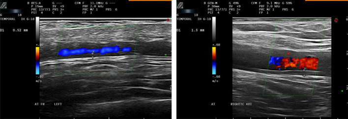
Retrospective observational study including patients referred to an US fast-track clinic. All patients underwent bilateral US examination of the cranial and extracranial arteries including the IMT measurement. Clinical confirmation of GCA after 6 months was considered the gold standard for diagnosis. A receiver operating characteristic (ROC) analysis was performed to select the cut-off values on the basis of the best tradeoff values between sensitivity and specificity.

RESULTS

A total of 157 patients were included, 47 (29.9%) with clinical confirmation of GCA after 6 months. 41 (87.2%) of patients with GCA had positive US findings (61.7% had cranial and 44.7% extracranial involvement). The best threshold IMT values were 0.44 mm for the common temporal artery; 0.34 mm for the frontal branch; 0.36 mm for the parietal branch; 1.1 mm for the carotid artery and 1 mm for the subclavian and axillary arteries. The areas under the ROC curves were greater for axillary arteries 0.996 (95% CI 0.991-1), for parietal branch 0.991 (95% CI 0.980-1), for subclavian 0.990 (95% CI 0.979-1), for frontal branch 0.989 (95% CI 0.976-1), for common temporal artery 0.984 (95% CI 0.959-1) and for common carotid arteries 0.977 (95% CI 0.961-0.993).

CONCLUSION

IMT cut-off values have been identified for each artery. These proposed IMT cut-off values may help to improve the diagnostic accuracy of US in clinical practice.

摘要

目的

确定颅内外动脉内膜中层厚度(IMT)的最佳超声(US)临界值,以鉴别巨细胞动脉炎(GCA)患者与非GCA患者。

方法

一项回顾性观察研究,纳入到超声快速诊所就诊的患者。所有患者均接受颅内外动脉的双侧超声检查,包括IMT测量。6个月后GCA的临床确诊被视为诊断的金标准。进行了受试者操作特征(ROC)分析,以根据敏感性和特异性之间的最佳权衡值选择临界值。

结果

共纳入157例患者,47例(29.9%)在6个月后经临床确诊为GCA。41例(87.2%)GCA患者超声检查结果为阳性(61.7%有颅内受累,44.7%有颅外受累)。最佳临界IMT值分别为:颞浅动脉0.44mm;额支0.34mm;顶支0.36mm;颈动脉1.1mm;锁骨下动脉和腋动脉1mm。腋动脉的ROC曲线下面积为0.996(95%CI 0.991-1),顶支为0.991(95%CI 0.980-1),锁骨下动脉为0.990(95%CI 0.979-1),额支为0.989(95%CI 0.976-1),颞浅动脉为0.984(95%CI 0.959-1),颈总动脉为0.977(95%CI 0.961-0.993)。

结论

已确定各动脉的IMT临界值。这些提出的IMT临界值可能有助于提高超声在临床实践中的诊断准确性。

https://cdn.ncbi.nlm.nih.gov/pmc/blobs/fe8d/9459085/91ed1d573be2/fmed-09-981804-g001.jpg

相似文献

1
Ultrasound intima media thickness cut-off values for cranial and extracranial arteries in patients with suspected giant cell arteritis.疑似巨细胞动脉炎患者颅内外动脉的超声内膜中层厚度截断值
Front Med (Lausanne). 2022 Aug 26;9:981804. doi: 10.3389/fmed.2022.981804. eCollection 2022.
2
Intima-media thickness cut-off values depicting "halo sign" and potential confounder analysis for the best diagnosis of large vessel giant cell arteritis by ultrasonography.通过超声检查描绘“晕征”的内膜中层厚度截断值及对大血管巨细胞动脉炎最佳诊断的潜在混杂因素分析。
Front Med (Lausanne). 2022 Dec 13;9:1055524. doi: 10.3389/fmed.2022.1055524. eCollection 2022.
3
Ultrasound cut-off values for intima-media thickness of temporal, facial and axillary arteries in giant cell arteritis.颞动脉、面动脉和腋动脉内-中膜厚度的巨细胞动脉炎超声截断值。
Rheumatology (Oxford). 2017 Sep 1;56(9):1479-1483. doi: 10.1093/rheumatology/kex143.
4
The cut-off values for the intima-media complex thickness assessed by colour Doppler sonography in seven cranial and aortic arch arteries.彩色多普勒超声检查 7 条颅动脉和主动脉弓动脉内中膜复合体厚度的截断值。
Rheumatology (Oxford). 2021 Mar 2;60(3):1346-1352. doi: 10.1093/rheumatology/keaa578.
5
Diagnosing vasculitis with ultrasound: findings and pitfalls.超声诊断血管炎:发现与陷阱
Ther Adv Musculoskelet Dis. 2024 Jun 5;16:1759720X241251742. doi: 10.1177/1759720X241251742. eCollection 2024.
6
Diagnostic validity of ultrasound including extra-cranial arteries in giant cell arteritis.巨细胞动脉炎中颅外动脉超声检查的诊断价值。
Clin Rheumatol. 2023 Apr;42(4):1163-1169. doi: 10.1007/s10067-022-06420-8. Epub 2022 Nov 11.
7
Is the intima-media thickness of temporal and axillary arteries influenced by cardiovascular risk?颞动脉和腋动脉的内中膜厚度是否受心血管风险的影响?
Rheumatology (Oxford). 2021 Nov 3;60(11):5362-5368. doi: 10.1093/rheumatology/keab117.
8
Ultrasound halo count in the differential diagnosis of atherosclerosis and large vessel giant cell arteritis.超声晕征计数在动脉粥样硬化和巨细胞动脉炎大血管病变鉴别诊断中的应用。
Arthritis Res Ther. 2023 Feb 14;25(1):23. doi: 10.1186/s13075-023-03002-0.
9
B-mode sonography wall thickness assessment of the temporal and axillary arteries for the diagnosis of giant cell arteritis: a cohort study.用于诊断巨细胞动脉炎的颞动脉和腋动脉B型超声检查壁厚评估:一项队列研究
Clin Exp Rheumatol. 2017 Mar-Apr;35 Suppl 103(1):128-133. Epub 2017 Apr 4.
10
The Usefullness of Subclavian Artery Ultrasound Assessment in Giant Cell Arteritis Evaluation.锁骨下动脉超声评估在巨细胞动脉炎评估中的实用性
J Clin Rheumatol. 2023 Jan 1;29(1):43-46. doi: 10.1097/RHU.0000000000001909. Epub 2022 Sep 20.

引用本文的文献

1
2D-cranial T1-black-blood MRI in suspected giant cell arteritis-measurement of vessel wall thickness does not give a diagnostic advantage compared to visual scoring alone.疑似巨细胞动脉炎患者的二维头颅T1加权黑血磁共振成像——与单纯视觉评分相比,血管壁厚度测量并无诊断优势。
Front Radiol. 2025 Jul 1;5:1597938. doi: 10.3389/fradi.2025.1597938. eCollection 2025.
2
OMERACT GCA phantom project: validation of a 3D-printed ultrasound training phantom for diagnosis of giant cell arteritis.OMERACT 巨细胞动脉炎模型项目:用于巨细胞动脉炎诊断的 3D 打印超声训练模型的验证
RMD Open. 2025 Feb 6;11(1):e005316. doi: 10.1136/rmdopen-2024-005316.
3

本文引用的文献

1
The Diagnostic Performance of an Extended Ultrasound Protocol in Patients With Clinically Suspected Giant Cell Arteritis.扩展超声方案对临床疑似巨细胞动脉炎患者的诊断效能
Front Med (Lausanne). 2022 Jan 18;8:807996. doi: 10.3389/fmed.2021.807996. eCollection 2021.
2
Subclavian artery involvement in patients with giant cell arteritis: do we need a modified Halo Score?巨细胞动脉炎患者锁骨下动脉受累:我们是否需要改良的 Halo 评分?
Clin Rheumatol. 2021 Jul;40(7):2821-2827. doi: 10.1007/s10067-020-05577-4. Epub 2021 Jan 11.
3
The cut-off values for the intima-media complex thickness assessed by colour Doppler sonography in seven cranial and aortic arch arteries.
Editorial: New insights into the role of imaging in large vessel vasculitis.
社论:影像学在大血管血管炎中作用的新见解
Front Med (Lausanne). 2024 Dec 3;11:1528452. doi: 10.3389/fmed.2024.1528452. eCollection 2024.
4
Supra-aortal intima-media thickness in treatment-naïve polymyalgia patients compared to matched controls.初治多肌痛患者与匹配对照组相比的主动脉弓内膜中层厚度。
Clin Rheumatol. 2024 Aug;43(8):2647-2651. doi: 10.1007/s10067-024-07021-3. Epub 2024 Jun 22.
5
Diagnosing vasculitis with ultrasound: findings and pitfalls.超声诊断血管炎:发现与陷阱
Ther Adv Musculoskelet Dis. 2024 Jun 5;16:1759720X241251742. doi: 10.1177/1759720X241251742. eCollection 2024.
6
Ultrasound intima-media thickness cut-off values for the diagnosis of giant cell arteritis using a dual clinical and MRI reference standard and cardiovascular risk stratification.采用临床和MRI双重参考标准及心血管风险分层诊断巨细胞动脉炎的超声内膜中层厚度临界值
Front Med (Lausanne). 2024 Apr 9;11:1389655. doi: 10.3389/fmed.2024.1389655. eCollection 2024.
7
Increased vertebral canal diameter measured by ultrasonography as a sign of vasculitis in patients with giant cell arteritis.超声测量显示巨细胞动脉炎患者椎管直径增加,作为血管炎的一个体征。
Front Med (Lausanne). 2023 Nov 7;10:1283285. doi: 10.3389/fmed.2023.1283285. eCollection 2023.
8
[Imaging of large vessel vasculitis].[大血管血管炎的影像学]
Z Rheumatol. 2023 Oct;82(8):646-653. doi: 10.1007/s00393-023-01405-7. Epub 2023 Aug 24.
9
Performance of the 2022 ACR/EULAR giant cell arteritis classification criteria for diagnosis in patients with suspected giant cell arteritis in routine clinical care.2022 年 ACR/EULAR 巨细胞动脉炎分类标准在常规临床护理中诊断疑似巨细胞动脉炎患者的性能。
RMD Open. 2023 Apr;9(2). doi: 10.1136/rmdopen-2022-002970.
10
Ultrasound halo count in the differential diagnosis of atherosclerosis and large vessel giant cell arteritis.超声晕征计数在动脉粥样硬化和巨细胞动脉炎大血管病变鉴别诊断中的应用。
Arthritis Res Ther. 2023 Feb 14;25(1):23. doi: 10.1186/s13075-023-03002-0.
彩色多普勒超声检查 7 条颅动脉和主动脉弓动脉内中膜复合体厚度的截断值。
Rheumatology (Oxford). 2021 Mar 2;60(3):1346-1352. doi: 10.1093/rheumatology/keaa578.
4
False positives in the ultrasound diagnosis of giant cell arteritis: some diseases can also show the halo sign.巨细胞动脉炎的超声诊断假阳性:有些疾病也可表现出晕环征。
Rheumatology (Oxford). 2020 Sep 1;59(9):2443-2447. doi: 10.1093/rheumatology/kez641.
5
Positive ultrasound halo sign of temporal arteries due to amyloidosis.颞动脉淀粉样变性的超声阳性晕征。
Rheumatology (Oxford). 2019 Nov 1;58(11):2067-2069. doi: 10.1093/rheumatology/kez182.
6
Diagnostic performance of temporal artery ultrasound for the diagnosis of giant cell arteritis: a systematic review and meta-analysis of the literature.颞动脉超声对巨细胞动脉炎诊断的诊断性能:文献的系统评价和荟萃分析。
Autoimmun Rev. 2019 Jan;18(1):56-61. doi: 10.1016/j.autrev.2018.07.012. Epub 2018 Nov 5.
7
Ultrasound in the diagnosis and management of giant cell arteritis.超声在巨细胞动脉炎的诊断和治疗中的应用。
Rheumatology (Oxford). 2018 Feb 1;57(suppl_2):ii22-ii31. doi: 10.1093/rheumatology/kex461.
8
Large-vessel giant cell arteritis: diagnosis, monitoring and management.巨细胞动脉炎(大动脉炎):诊断、监测和管理。
Rheumatology (Oxford). 2018 Feb 1;57(suppl_2):ii32-ii42. doi: 10.1093/rheumatology/kex424.
9
Definitions and reliability assessment of elementary ultrasound lesions in giant cell arteritis: a study from the OMERACT Large Vessel Vasculitis Ultrasound Working Group.巨细胞动脉炎中基本超声病变的定义及可靠性评估:来自OMERACT大血管血管炎超声工作组的一项研究
RMD Open. 2018 May 17;4(1):e000598. doi: 10.1136/rmdopen-2017-000598. eCollection 2018.
10
Imaging in diagnosis, outcome prediction and monitoring of large vessel vasculitis: a systematic literature review and meta-analysis informing the EULAR recommendations.大血管血管炎诊断、预后预测及监测中的影像学检查:一项为欧洲抗风湿病联盟(EULAR)推荐意见提供依据的系统文献综述和荟萃分析
RMD Open. 2018 Feb 2;4(1):e000612. doi: 10.1136/rmdopen-2017-000612. eCollection 2018.