Department of Electronic and Information Engineering, Harbin Institute of Technology at Shenzhen, Shenzhen, China.
Peng Cheng Laboratory, Shenzhen, China.
Hum Brain Mapp. 2022 Nov;43(16):5017-5031. doi: 10.1002/hbm.26066. Epub 2022 Sep 12.
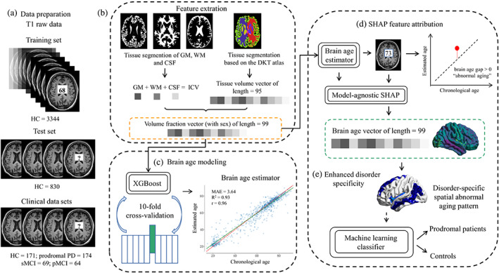
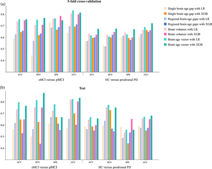
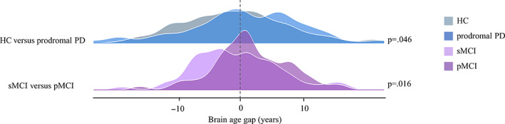
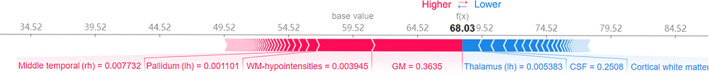
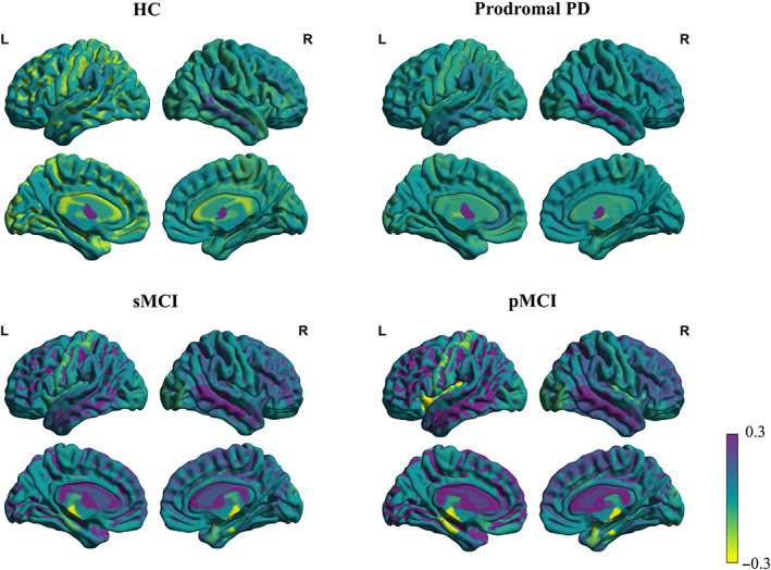
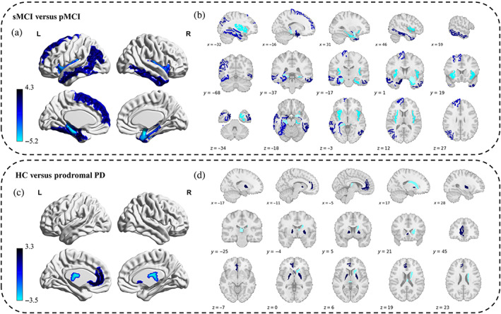
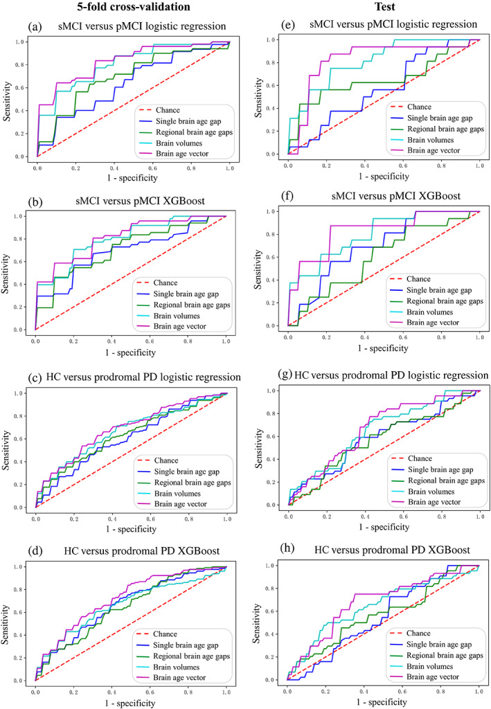
Neuroimaging-driven brain age estimation has become popular in measuring brain aging and identifying neurodegenerations. However, the single estimated brain age (gap) compromises regional variations of brain aging, losing spatial specificity across diseases which is valuable for early screening. In this study, we combined brain age modeling with Shapley Additive Explanations to measure brain aging as a feature contribution vector underlying spatial pathological aging mechanism. Specifically, we regressed age with volumetric brain features using machine learning to construct the brain age model, and model-agnostic Shapley values were calculated to attribute regional brain aging for each subject's age estimation, forming the brain age vector. Spatial specificity of the brain age vector was evaluated among groups of normal aging, prodromal Parkinson disease (PD), stable mild cognitive impairment (sMCI), and progressive mild cognitive impairment (pMCI). Machine learning methods were adopted to examine the discriminability of the brain age vector in early disease screening, compared with the other two brain aging metrics (single brain age gap, regional brain age gaps) and brain volumes. Results showed that the proposed brain age vector accurately reflected disorder-specific abnormal aging patterns related to the medial temporal and the striatum for prodromal AD (sMCI vs. pMCI) and PD (healthy controls [HC] vs. prodromal PD), respectively, and demonstrated outstanding performance in early disease screening, with area under the curves of 83.39% and 72.28% in detecting pMCI and prodromal PD, respectively. In conclusion, the proposed brain age vector effectively improves spatial specificity of brain aging measurement and enables individual screening of neurodegenerative diseases.
神经影像学驱动的大脑年龄估计在衡量大脑衰老和识别神经退行性变方面变得流行。然而,单一估计的大脑年龄(差距)损害了大脑衰老的区域变化,丧失了疾病之间具有价值的早期筛查的空间特异性。在这项研究中,我们将大脑年龄建模与 Shapley 加法解释相结合,以测量大脑衰老作为潜在的空间病理衰老机制的特征贡献向量。具体来说,我们使用机器学习回归年龄与容积脑特征,构建大脑年龄模型,并计算模型不可知的 Shapley 值,以将区域大脑衰老归因于每个受试者的年龄估计,形成大脑年龄向量。在正常衰老、前驱帕金森病(PD)、稳定轻度认知障碍(sMCI)和进行性轻度认知障碍(pMCI)的组中评估大脑年龄向量的空间特异性。采用机器学习方法检查大脑年龄向量在早期疾病筛查中的可区分性,与其他两种大脑老化指标(单一大脑年龄差距、区域大脑年龄差距)和脑体积相比。结果表明,所提出的大脑年龄向量准确地反映了与前驱 AD(sMCI 与 pMCI)和 PD(健康对照 [HC] 与前驱 PD)相关的内侧颞叶和纹状体的特定疾病异常衰老模式,并且在早期疾病筛查中表现出色,在检测 pMCI 和前驱 PD 时的曲线下面积分别为 83.39%和 72.28%。总之,所提出的大脑年龄向量有效地提高了大脑衰老测量的空间特异性,并实现了神经退行性疾病的个体筛查。