Department of Ophthalmology, University of California Irvine, Gavin Herbert Eye Institute, Irvine, CA, USA.
Northwest Eye Surgeons, Seattle, WA, USA.
Indian J Ophthalmol. 2022 Oct;70(10):3669-3672. doi: 10.4103/ijo.IJO_391_22.
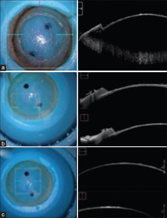
We aimed to develop a novel and effective technique for creating a smooth deep lamellar dissection of the cornea using a femtosecond (FS) laser for deep anterior lamellar keratoplasty (DALK), we conducted a retrospective eye bank study. Thirteen fresh human corneas were mounted on an artificial anterior chamber, and deep lamellar cuts were made with a 500-kHz VisuMax FS laser at a level of 50-80 μm anterior to the Descemet's membrane (DM). A posterior diameter of 8 mm with a side cut angle of 110° was used for the anterior penetrating side cut. The anterior lamellar tissue was bluntly dissected. The residual posterior stromal beds and side cuts were examined with microscopy and intraoperative optical coherence tomography (OCT) and post-cut endothelial cell evaluations. All corneas revealed a smooth residual posterior stromal bed without any visible irregularities or ridges by microscopy and OCT imaging. Six corneas were suitable for post-cut endothelial cell evaluation 2 days after laser cut, with no significant endothelial cell loss post-laser and blunt dissection of the posterior stroma. FS laser deep lamellar keratoplasty utilizing an ultrafast laser to produce a smooth deep stromal dissection followed by blunt dissection and removal of the anterior stromal tissue yields a consistent and smooth residual stromal bed. The creation of a smooth lamellar dissection in the deep posterior cornea may result in more consistent DALK without the need for air bubble or manual baring of DM that has the risk for DM perforation.
我们旨在开发一种新的有效技术,使用飞秒(FS)激光对角膜进行深层板层解剖,以进行深前板层角膜移植术(DALK),我们进行了一项回顾性眼库研究。将 13 个新鲜的人眼角膜安装在人工前房上,并使用 500-kHz VisuMax FS 激光在距 Descemet 膜(DM)前 50-80μm 的水平进行深层板层切割。用于前穿透侧切口的后侧直径为 8mm,侧切口角度为 110°。前板层组织被钝性解剖。使用显微镜和术中光学相干断层扫描(OCT)以及切割后内皮细胞评估来检查残留的后基质床和侧切口。所有角膜在显微镜和 OCT 成像下均显示出光滑的残留后基质床,没有任何可见的不规则或脊。六只眼角膜在激光切割后 2 天适合进行切割后内皮细胞评估,激光切割后和后基质钝性解剖后内皮细胞无明显丢失。利用超快激光制作光滑深层基质解剖,然后进行钝性解剖和前基质组织切除的 FS 激光深层板层角膜移植术可产生一致且光滑的残留基质床。在深层后角膜中制作光滑的板层解剖可能会导致更一致的 DALK,而无需气泡或手动暴露 DM,因为这有 DM 穿孔的风险。