Guangdong Eye Institute, Department of Ophthalmology, Guangdong Provincial People's Hospital, Guangdong Academy of Medical Sciences, Guangzhou, China.
Guangdong Cardiovascular Institute, Guangdong Provincial People's Hospital, Guangdong Academy of Medical Sciences, Guangzhou, China.
JAMA Netw Open. 2022 Oct 3;5(10):e2235017. doi: 10.1001/jamanetworkopen.2022.35017.
Vision loss and depression are common conditions with major health implications. However, mechanisms of the association of visual health (across the full acuity spectrum) with depression remain unclear.
To characterize the association between visual health and depression and investigate the association between depression and brain microstructure and macrostructure in subgroups divided by visual acuity.
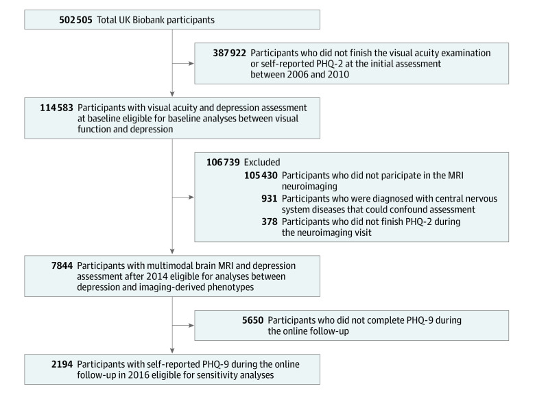
DESIGN, SETTING, AND PARTICIPANTS: In the UK Biobank Study cohort, 114 583 volunteers were included at baseline from March to June 2006 to July 2010. Habitual distance visual acuity was examined using the logarithm of the minimum angle of resolution (LogMAR) characters. Depression was identified based on Patient Health Questionnaire (PHQ) or through an interview-based psychiatric diagnosis. Subgroup participants completed multimodal magnetic resonance imaging (MRI) of the brain and PHQ evaluation during the imaging visit after 2014. Data were analyzed from May 5 to August 9, 2022.
Depression, depressive symptoms, and imaging-derived phenotypes from T1-weighted and diffusion MRI.
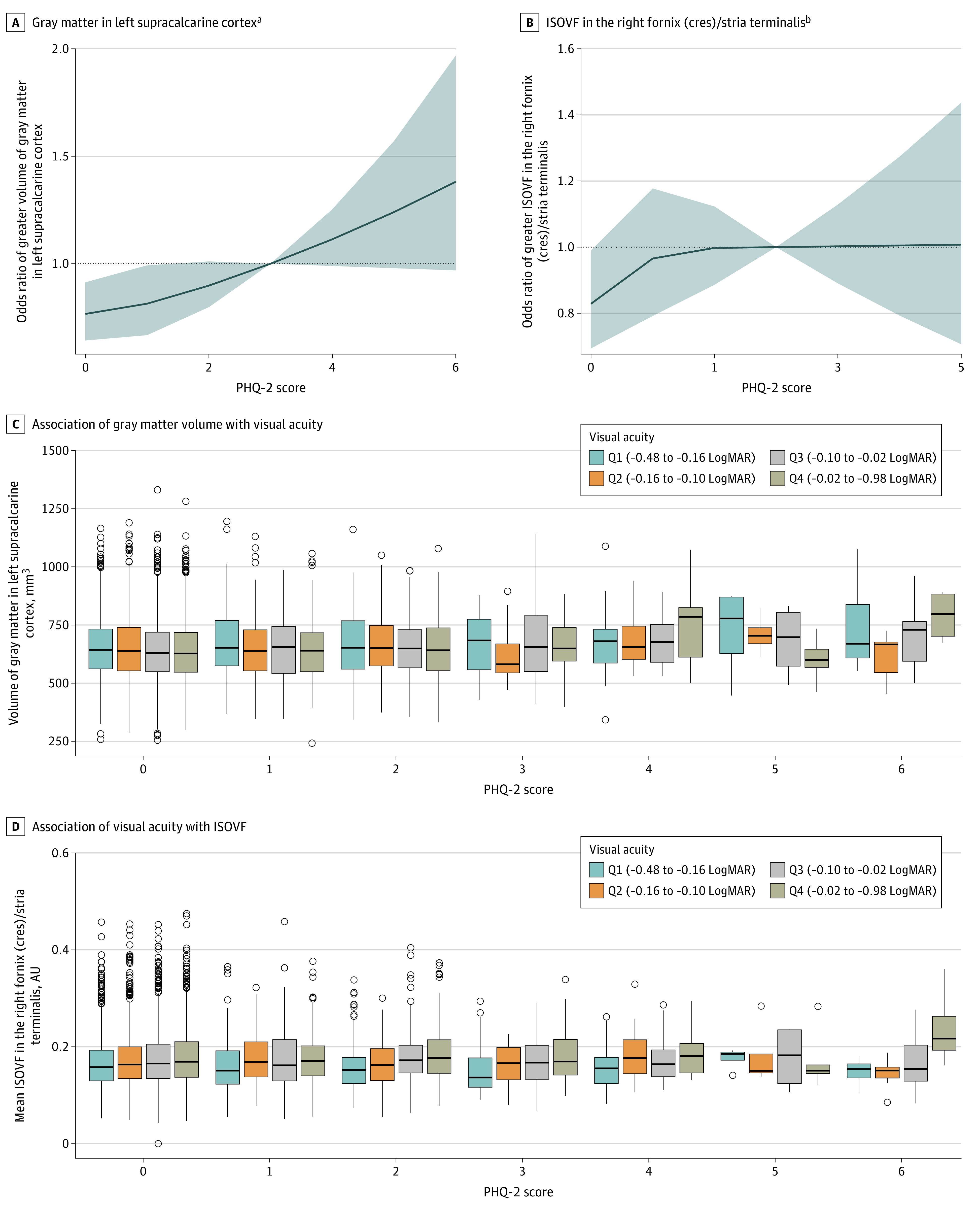
Of the 114 583 participants from the UK Biobank Study, 62 401 (54.5%) were women, and the mean (SD) age was 56.8 (8.1) years (range, 39-72 years). A 1-line worse visual acuity (0.1 LogMAR increase) was associated with 5% higher odds of depression (odds ratio, 1.05 [95% CI, 1.04-1.07]) after adjustment for age, sex, race and ethnicity, Townsend index, educational qualifications, smoking, alcohol consumption, obesity, physical activity, history of hypertension, diabetes, hyperlipidemia, and family history of depression. Of the 7844 participants eligible for MRI analysis, there were linear associations between PHQ score and the left volume of gray matter in supracalcarine cortex (coefficient, 7.61 [95% CI, 3.90-11.31]) and mean isotropic volume fraction (ISOVF) in the right fornix (cres) and/or stria terminalis (coefficient, 0.003 [95% CI, 0.001-0.004]) after correction for multiple comparison. In addition, their association could be moderated by visual acuity, whereby increased PHQ score was associated with higher ISOVF levels only among those with poorer visual acuity (P = .02 for interaction).
This study suggests an association between visual health and depression and that the diffusion characteristic of ISOVF in the fornix (cres) and/or stria terminalis is associated with depressive symptoms in participants with poorer visual acuity.
视力丧失和抑郁是常见的病症,对健康有重大影响。然而,视觉健康(全视力范围)与抑郁之间的关联机制仍不清楚。
描述视觉健康与抑郁之间的关联,并在根据视力分组的亚组中调查抑郁与大脑微观结构和宏观结构之间的关联。
设计、地点和参与者:在英国生物库研究队列中,共有 114583 名志愿者于 2006 年 3 月至 6 月至 2010 年 7 月基线时纳入研究。使用最小角分辨率(LogMAR)字符检查习惯性远距离视力。抑郁通过患者健康问卷(PHQ)或基于访谈的精神病学诊断确定。亚组参与者在 2014 年后的成像访问期间完成了大脑的多模态磁共振成像(MRI)和 PHQ 评估。数据于 2022 年 5 月 5 日至 8 月 9 日进行分析。
抑郁、抑郁症状和来自 T1 加权和弥散 MRI 的成像衍生表型。
在英国生物库研究中的 114583 名参与者中,有 62401 名(54.5%)为女性,平均(SD)年龄为 56.8(8.1)岁(范围 39-72 岁)。视力每下降一行(0.1 LogMAR 增加),抑郁的几率就会增加 5%(优势比,1.05 [95%CI,1.04-1.07]),调整年龄、性别、种族和民族、汤森指数、教育程度、吸烟、饮酒、肥胖、身体活动、高血压史、糖尿病、高脂血症和抑郁家族史后。在符合 MRI 分析条件的 7844 名参与者中,PHQ 评分与左侧超距皮质灰质体积(系数,7.61 [95%CI,3.90-11.31])和右侧穹窿(cres)和/或终纹平均各向同性体积分数(ISOVF)呈线性关联(系数,0.003 [95%CI,0.001-0.004]),校正多重比较后。此外,他们的关联可以通过视力来调节,即仅在视力较差的参与者中,PHQ 评分的增加与更高的 ISOVF 水平相关(P=0.02 交互作用)。
本研究表明视觉健康与抑郁之间存在关联,穹窿(cres)和/或终纹的 ISOVF 弥散特征与视力较差参与者的抑郁症状相关。