College of Medicine and Life Sciences, University of Toledo, Toledo, OH, USA.
Department of Medical Education/Neurosciences, College of Medicine and Life Sciences University of Toledo, Toledo, OH, USA.
Am J Case Rep. 2022 Oct 11;23:e937450. doi: 10.12659/AJCR.937450.
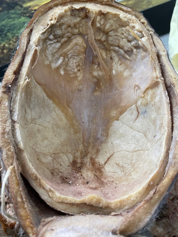
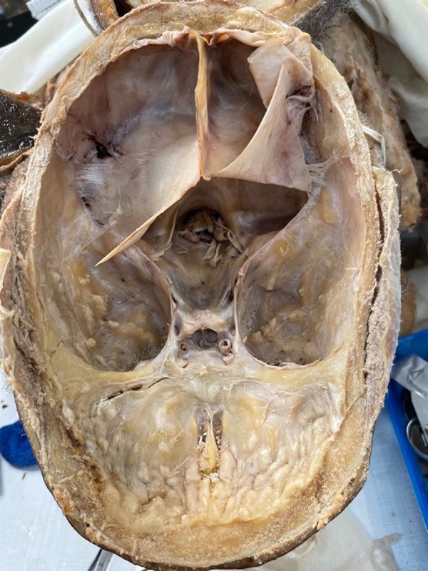
BACKGROUND Hyperostosis frontalis interna is a boney overgrowth of the inner side of the frontal bone of the skull caused by overgrowth of the endocranial surface. It is most often found in women after menopause. It is also associated with hormonal imbalance, being overweight, history of headaches, and neurocognitive degenerative conditions. Female gender, advanced age, extended estrogen stimulation, and elevated leptin levels may also play a role. The thickening is usually confined to the frontal bone, but it can spread as far as the anterior parietal and temporal bones. CASE REPORT During a medical school dissection course, an extensive boney overgrowth in the frontal regions covering the inside of the frontal bone of the skull of a 90-year-old female donor, who died of a cerebrovascular infarction, was identified. This boney overgrowth was mainly confined within the frontal region, but there was some boney overgrowth that extended to the temporal bones. The overgrowth in the endocranium of the temporal bone was not as severe as the overgrowth of the frontal bone. The morphology of the overgrowth was rigid, uneven, and bumpy. Based upon the physical characteristics, we concluded that this presentation was consistent with hyperostosis frontalis interna. CONCLUSIONS Our female donor was found to exhibit a phenomenon which could be clinically underdiagnosed due to its internal nature and asymptomatic presentation. Insight into the potential causes of HFI and its identification during clinical evaluation offers a path for future research to better identify and manage cases of HFI.
颅腔内部额骨过度生长是颅骨额骨内侧的骨质过度生长,由颅腔内部表面过度生长引起。它最常发生在绝经后的女性中。它还与激素失衡、超重、头痛史和神经认知退行性疾病有关。女性、高龄、延长的雌激素刺激和升高的瘦素水平也可能起作用。增厚通常局限于额骨,但也可能扩散到额骨和颞骨的前部。
在医学院解剖课程中,发现一名 90 岁女性捐献者颅骨额骨内侧有广泛的骨质过度生长,该捐献者死于脑血管梗塞。这种骨质过度生长主要局限于额骨区域,但也有一些骨质过度生长延伸至颞骨。颞骨颅腔内部的过度生长不如额骨严重。过度生长的形态坚硬、不均匀且凹凸不平。根据其物理特征,我们得出结论,这种表现与颅腔内部额骨过度生长一致。
我们的女性捐献者表现出一种可能由于其内部性质和无症状表现而被临床误诊的现象。深入了解 HFI 的潜在原因及其在临床评估中的识别为未来的研究提供了一条途径,以更好地识别和管理 HFI 病例。