Department of Breast Surgery, First Affiliated Hospital of Xi'an Jiaotong University, Xi'an, China.
Department of Structural Heart Disease, First Affiliated Hospital of Xi'an Jiaotong University, Xi'an, China.
Anal Cell Pathol (Amst). 2022 Oct 3;2022:5942379. doi: 10.1155/2022/5942379. eCollection 2022.
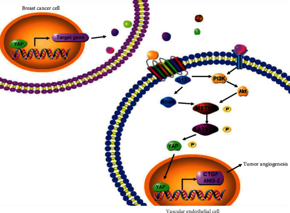
The YAP signaling pathway is altered and implicated as oncogenic in human mammary cancers. However, roles of YAP signaling that regulate the breast tumor angiogenesis have remained elusive. Tumor angiogenesis is coordinated by the activation of both cancer cells and vascular endothelial cells. Whether the YAP signaling pathway can regulate the intercellular interaction between cancer cells and endothelial cells is essentially unknown.
The effects of YAP on tumor angiogenesis, migration, and proliferation of vascular endothelial cells were evaluated in vitro. Expression of proteins and phosphorylating proteins involved in YAP, G13-RhoA, and PI3K/Akt signaling pathways was evaluated using the Western blotting, immunofluorescence staining, and immunohistochemistry analysis. In addition, the effects of YAP on breast cancer angiogenesis were evaluated in vivo by tumor xenograft mice.
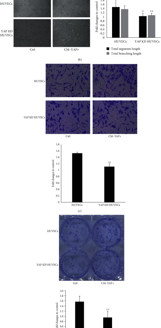
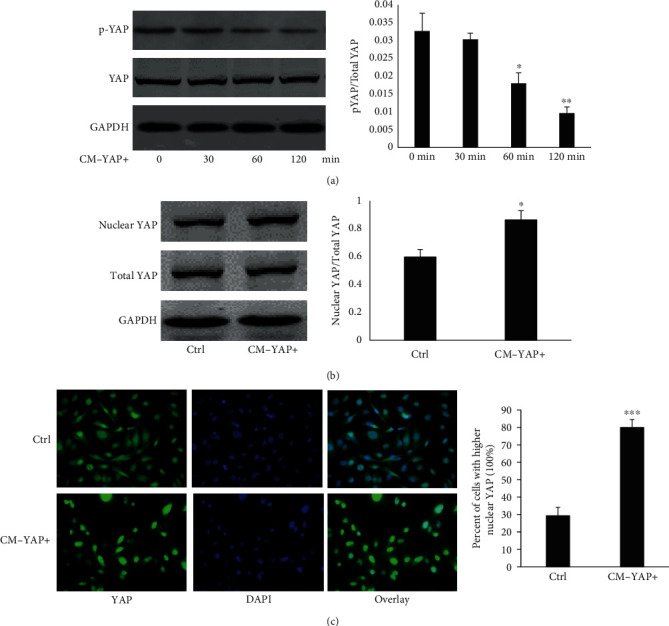
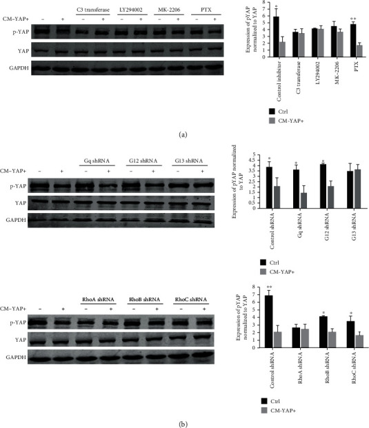
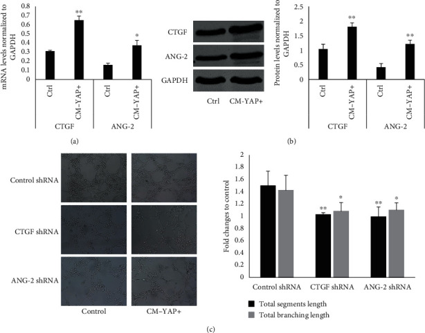
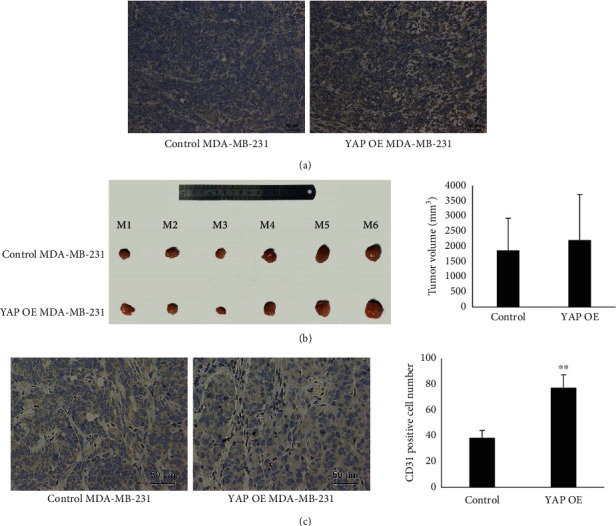
We showed here that conditioned media from YAP overexpressed breast cancer cells (CM-YAP+) could promote angiogenesis, accompanied by increased tube formation, migration, and proliferation of human umbilical vein endothelial cells (HUVECs). Down regulation of YAP in HUVECs reversed CM-YAP+ induced angiogenesis. CM-YAP+ time-dependently activated YAP in HUVECs by dephosphorylating YAP and increasing nuclear translocation. We also identified that both G-RhoA and PI3K/Akt signaling pathway were necessary for CM-YAP+ induced activation of YAP. Besides, connective tissue growth factor (CTGF) and angiopoietin-2 (ANG-2) acted as down-stream of YAP in HUVECs to promote angiogenesis. In addition, subcutaneous tumors nude mice model demonstrated that tumors overexpressed YAP revealed more neovascularization in vivo.
YAP-YAP interaction between breast cancer cells and endothelial cells could promote tumor angiogenesis, supporting that YAP is a potential marker and target for developing novel therapeutic strategies against breast cancer.
YAP 信号通路在人类乳腺癌中发生改变并被认为具有致癌作用。然而,YAP 信号调节乳腺肿瘤血管生成的作用仍不清楚。肿瘤血管生成由癌细胞和血管内皮细胞的激活共同协调。YAP 信号通路是否可以调节癌细胞和内皮细胞之间的细胞间相互作用在本质上尚不清楚。
在体外评估 YAP 对肿瘤血管生成、血管内皮细胞迁移和增殖的影响。使用 Western blot、免疫荧光染色和免疫组织化学分析评估涉及 YAP、G13-RhoA 和 PI3K/Akt 信号通路的蛋白质和磷酸化蛋白质的表达。此外,通过肿瘤异种移植小鼠评估 YAP 对乳腺癌血管生成的影响。
我们在这里表明,YAP 过表达乳腺癌细胞的条件培养基(CM-YAP+)可促进血管生成,伴随人脐静脉内皮细胞(HUVEC)的管形成、迁移和增殖增加。在 HUVEC 中下调 YAP 可逆转 CM-YAP+诱导的血管生成。CM-YAP+可通过去磷酸化 YAP 和增加核易位使 HUVEC 中的 YAP 时间依赖性地激活。我们还发现 G-RhoA 和 PI3K/Akt 信号通路对于 CM-YAP+诱导的 YAP 激活都是必要的。此外,结缔组织生长因子(CTGF)和血管生成素-2(ANG-2)在 HUVECs 中作为 YAP 的下游分子促进血管生成。此外,裸鼠皮下肿瘤模型表明,过表达 YAP 的肿瘤在体内显示出更多的新生血管化。
乳腺癌细胞和内皮细胞之间的 YAP-YAP 相互作用可促进肿瘤血管生成,这支持 YAP 是开发针对乳腺癌的新型治疗策略的潜在标志物和靶标。