• 文献检索
  • 文档翻译
  • 深度研究
  • 学术资讯
  • Suppr Zotero 插件Zotero 插件
  • 邀请有礼
  • 套餐&价格
  • 历史记录
应用&插件
Suppr Zotero 插件Zotero 插件浏览器插件Mac 客户端Windows 客户端微信小程序
定价
高级版会员购买积分包购买API积分包
服务
文献检索文档翻译深度研究API 文档MCP 服务
关于我们
关于 Suppr公司介绍联系我们用户协议隐私条款
关注我们

Suppr 超能文献

核心技术专利:CN118964589B侵权必究
粤ICP备2023148730 号-1Suppr @ 2026

文献检索

告别复杂PubMed语法,用中文像聊天一样搜索,搜遍4000万医学文献。AI智能推荐,让科研检索更轻松。

立即免费搜索

文件翻译

保留排版,准确专业,支持PDF/Word/PPT等文件格式,支持 12+语言互译。

免费翻译文档

深度研究

AI帮你快速写综述,25分钟生成高质量综述,智能提取关键信息,辅助科研写作。

立即免费体验

非平足和 II 期成人获得性平足在站立相期间的后足关节运动变化的比较。

Comparison of in vivo hindfoot joints motion changes during stance phase between non-flatfoot and stage II adult acquired flatfoot.

机构信息

Department of Sports Medicine, The First Affiliated Hospital of Shenzhen University, Shenzhen Second People's Hospital, Shenzhen, Guangdong, China.

Department of Orthopaedics, Xiangya Hospital, Central South University, Changsha, Hunan, China.

出版信息

J Foot Ankle Res. 2022 Oct 13;15(1):74. doi: 10.1186/s13047-022-00577-w.

DOI:10.1186/s13047-022-00577-w
PMID:36229819
原文链接:https://pmc.ncbi.nlm.nih.gov/articles/PMC9559000/
Abstract

BACKGROUND

To compare the kinematic characteristics of hindfoot joints in stage II adult acquired flatfoot deformity (AAFD) with those of non-flatfoot through the 3D-to-2D registration technology and single fluoroscopic imaging system.

METHODS

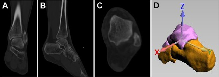
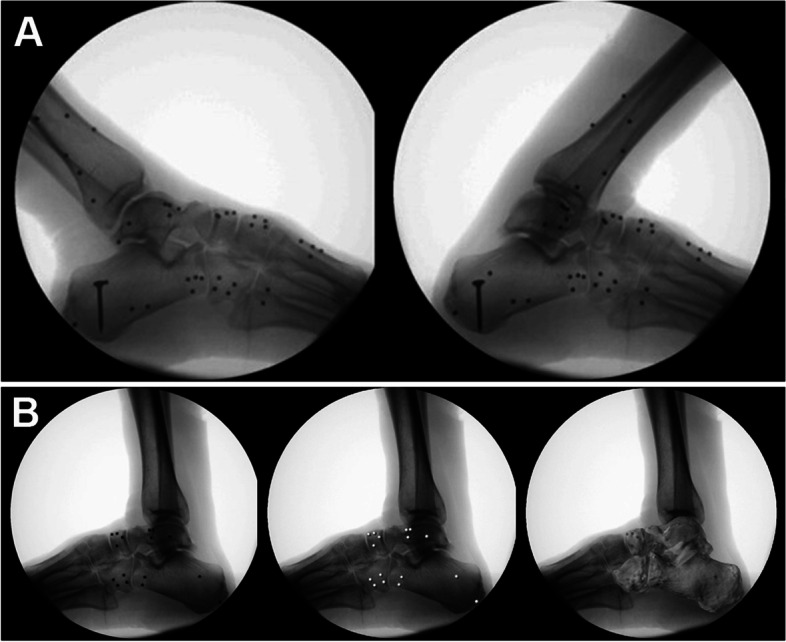
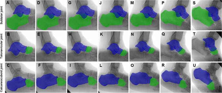
Eight volunteers with stage II AAFD and seven volunteers without stage II AAFD were recruited and CT scans were performed bilateral for both groups in neutral positions. Their lateral dynamic X-ray data during the stance phase, including 14 non-flatfeet and 10 flatfeet, was collected. A computer-aided simulated light source for 3D CT model was applied to obtain the virtual images, which were matched with the dynamic X-ray images to register in the "Fluo" software, so that the spatial changes during the stance phase could be calculated.

RESULTS

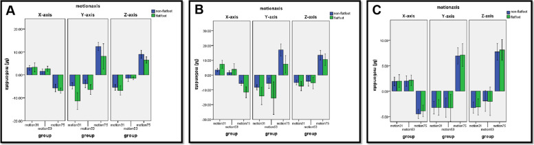
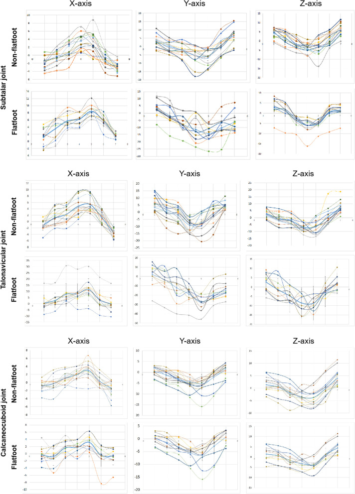
During the early-stance phase, the calcaneous was more dorsiflexed, everted, and externally-rotated relative to the talus in flatfoot compared with that in non-flatfoot (p < 0.05). During the mid-stance phase, the calcaneous was more dorsiflexed and everted relative to the talus in flatfoot compared with that in non-flatfoot (p < 0.05); however, the rotation did not differ significantly between the two groups (p > 0.05). During the late-stance phase, the calcaneous was more plantarflexed, but less inverted and internally-rotated, relative to the talus in flatfoot compared with that in non-flatfoot (p < 0.05). During the early- and mid-stance phase, the navicular was more dorsiflexed, everted, and externally-rotated relative to the talus in flatfoot compared with that in non-flatfoot (p < 0.05). During the late-stance phase, the navicular was more plantarflexed, but less inverted and internally-rotated, relative to the talus in flatfoot compared with that in non-flatfoot (p < 0.05). There was no difference in the motion of cuboid between the two groups during the whole stance phase (p > 0.05).

CONCLUSIONS

During the early- and mid-stance phase, excessive motion was observed in the subtalar and talonavicular joints in stage II AAFD. During the late-stance phase, the motion of subtalar and talonavicular joints appeared to be in the dysfunction state. The current study helps better understanding the biomechanics of the hindfoot during non-flatfoot and flatfoot condition which is critical to the intervention to the AAFD using conservative treatment such as insole or surgical treatment for joint hypermotion.

摘要

背景

通过三维到二维配准技术和单荧光透视成像系统,比较 II 期成人获得性平足畸形(AAFD)患者和非平足患者的后足关节运动学特征。

方法

招募 8 名 II 期 AAFD 志愿者和 7 名非 II 期 AAFD 志愿者,两组均在中立位进行双侧 CT 扫描。采集两组患者在站立相的动态侧位 X 线数据,共 14 个非平足和 10 个平足。应用计算机辅助模拟 3D CT 模型光源获得虚拟图像,将其与动态 X 线图像在“Fluo”软件中配准,以计算站立相时的空间变化。

结果

在早期站立相中,与非平足相比,平足患者距骨在背屈、外展和外旋方面更为明显(p<0.05)。在中期站立相中,与非平足相比,平足患者距骨在背屈和外展方面更为明显(p<0.05);然而,两组之间的旋转没有显著差异(p>0.05)。在晚期站立相中,与非平足相比,平足患者距骨的跖屈程度更大,但内翻和内旋程度更小(p<0.05)。在早期和中期站立相中,与非平足相比,平足患者的舟骨在背屈、外展和外旋方面更为明显(p<0.05)。在晚期站立相中,与非平足相比,平足患者的舟骨跖屈程度更大,但内翻和内旋程度更小(p<0.05)。在整个站立相中,两组患者的骰骨运动无差异(p>0.05)。

结论

在 II 期 AAFD 患者中,在早期和中期站立相中观察到距下和跟舟关节的过度运动。在晚期站立相中,距下和跟舟关节的运动似乎处于功能障碍状态。本研究有助于更好地了解非平足和平足状态下后足的生物力学特性,这对于使用鞋垫或关节过度活动的手术治疗等保守治疗方法干预 AAFD 至关重要。

https://cdn.ncbi.nlm.nih.gov/pmc/blobs/73aa/9559000/12d2279f0421/13047_2022_577_Fig7_HTML.jpg
https://cdn.ncbi.nlm.nih.gov/pmc/blobs/73aa/9559000/c3af033cf332/13047_2022_577_Fig1_HTML.jpg
https://cdn.ncbi.nlm.nih.gov/pmc/blobs/73aa/9559000/aff4d46caea1/13047_2022_577_Fig2_HTML.jpg
https://cdn.ncbi.nlm.nih.gov/pmc/blobs/73aa/9559000/d7e07743a36c/13047_2022_577_Fig3_HTML.jpg
https://cdn.ncbi.nlm.nih.gov/pmc/blobs/73aa/9559000/bc8a5e0c076f/13047_2022_577_Fig4_HTML.jpg
https://cdn.ncbi.nlm.nih.gov/pmc/blobs/73aa/9559000/fc11673b77c9/13047_2022_577_Fig5_HTML.jpg
https://cdn.ncbi.nlm.nih.gov/pmc/blobs/73aa/9559000/50e0700a9b72/13047_2022_577_Fig6_HTML.jpg
https://cdn.ncbi.nlm.nih.gov/pmc/blobs/73aa/9559000/12d2279f0421/13047_2022_577_Fig7_HTML.jpg

相似文献

1
Comparison of in vivo hindfoot joints motion changes during stance phase between non-flatfoot and stage II adult acquired flatfoot.非平足和 II 期成人获得性平足在站立相期间的后足关节运动变化的比较。
J Foot Ankle Res. 2022 Oct 13;15(1):74. doi: 10.1186/s13047-022-00577-w.
2
Pathological kinematic patterns of the tarsal complex in stage II adult-acquired flatfoot deformity.第二期成人获得性平足畸形中跗骨复合体的病理性运动模式。
J Orthop Res. 2019 Feb;37(2):477-482. doi: 10.1002/jor.23821. Epub 2019 Feb 1.
3
An in vivo study of hindfoot 3D kinetics in stage II posterior tibial tendon dysfunction (PTTD) flatfoot based on weight-bearing CT scan.基于负重 CT 扫描的 II 期跟骨后足三维动力学在胫骨后肌腱功能障碍(PTTD)平足中的体内研究。
Bone Joint Res. 2013 Dec 9;2(12):255-63. doi: 10.1302/2046-3758.212.2000220. Print 2013.
4
Three-dimensional kinematic change of hindfoot during full weightbearing in standing: an analysis using upright computed tomography and 3D-3D surface registration.站立位全负重时后足的三维运动学变化:一项使用直立计算机断层扫描和三维-三维表面配准的分析
J Orthop Surg Res. 2019 Nov 11;14(1):355. doi: 10.1186/s13018-019-1443-z.
5
Correlation between hindfoot joint three-dimensional kinematics and the changes of the medial arch angle in stage II posterior tibial tendon dysfunction flatfoot.Ⅱ期胫后肌腱功能不全扁平足中后足关节三维运动学与内侧足弓角度变化的相关性
Clin Biomech (Bristol). 2015 Feb;30(2):153-8. doi: 10.1016/j.clinbiomech.2014.12.007. Epub 2014 Dec 24.
6
Correlation of Talar Anatomy and Subtalar Joint Alignment on Weightbearing Computed Tomography With Radiographic Flatfoot Parameters.负重计算机断层扫描下距骨解剖结构及距下关节对线与平足症影像学参数的相关性
Foot Ankle Int. 2016 Aug;37(8):874-81. doi: 10.1177/1071100716646629. Epub 2016 May 2.
7
Load response of the tarsal bones in patients with flatfoot deformity: in vivo 3D study.足弓塌陷患者跗骨的负荷反应:体内 3D 研究。
Foot Ankle Int. 2011 Nov;32(11):1017-22. doi: 10.3113/FAI.2011.1017.
8
In vivo three-dimensional analysis of hindfoot kinematics in stage II PTTD flatfoot.II期胫后肌腱功能不全扁平足中后足运动学的体内三维分析
J Orthop Sci. 2015 May;20(3):488-97. doi: 10.1007/s00776-015-0698-4. Epub 2015 Feb 10.
9
Evaluation of peritalar subluxation in adult acquired flatfoot deformity using computed tomography and weightbearing multiplanar imaging.使用计算机断层扫描和负重多平面成像评估成人获得性平足畸形中的距周半脱位
Foot Ankle Surg. 2018 Dec;24(6):495-500. doi: 10.1016/j.fas.2017.05.010. Epub 2017 Jun 12.
10
Load response of the medial longitudinal arch in patients with flatfoot deformity: in vivo 3D study.扁平足畸形患者内侧纵弓的负荷反应:一项体内三维研究。
Clin Biomech (Bristol). 2013 Jun;28(5):568-73. doi: 10.1016/j.clinbiomech.2013.04.004. Epub 2013 May 2.

本文引用的文献

1
Adult Acquired Flatfoot Deformity: Anatomy, Biomechanics, Staging, and Imaging Findings.成人获得性平足畸形:解剖、生物力学、分期和影像学表现。
Radiographics. 2019 Sep-Oct;39(5):1437-1460. doi: 10.1148/rg.2019190046.
2
Flatfoot deformity affected the kinematics of the foot and ankle in proportion to the severity of deformity.平足畸形会根据畸形的严重程度,对足部和踝关节的运动学产生相应的影响。
Gait Posture. 2019 Jul;72:123-128. doi: 10.1016/j.gaitpost.2019.06.002. Epub 2019 Jun 5.
3
Analysis of biomechanical stresses caused by hindfoot joint arthrodesis in the treatment of adult acquired flatfoot deformity: A finite element study.
分析距下关节融合术治疗成人获得性平足畸形的生物力学应力:有限元研究。
Foot Ankle Surg. 2020 Jun;26(4):412-420. doi: 10.1016/j.fas.2019.05.010. Epub 2019 May 19.
4
Pathological kinematic patterns of the tarsal complex in stage II adult-acquired flatfoot deformity.第二期成人获得性平足畸形中跗骨复合体的病理性运动模式。
J Orthop Res. 2019 Feb;37(2):477-482. doi: 10.1002/jor.23821. Epub 2019 Feb 1.
5
Foot segmental motion and coupling in stage II and III tibialis posterior tendon dysfunction.Ⅱ期和Ⅲ期胫后肌腱功能障碍时足部节段性运动及耦合情况
Clin Biomech (Bristol). 2017 Jun;45:38-42. doi: 10.1016/j.clinbiomech.2017.04.007. Epub 2017 Apr 21.
6
Biplane fluoroscopy for hindfoot motion analysis during gait: A model-based evaluation.用于步态中后足运动分析的双平面荧光透视法:基于模型的评估
Med Eng Phys. 2017 May;43:118-123. doi: 10.1016/j.medengphy.2017.02.009. Epub 2017 Mar 1.
7
Outcomes of a Stepcut Lengthening Calcaneal Osteotomy for Adult-Acquired Flatfoot Deformity.成人获得性平足畸形的阶梯状延长跟骨截骨术的疗效
Foot Ankle Int. 2015 Jul;36(7):749-55. doi: 10.1177/1071100715574933. Epub 2015 Mar 2.
8
In vivo three-dimensional analysis of hindfoot kinematics in stage II PTTD flatfoot.II期胫后肌腱功能不全扁平足中后足运动学的体内三维分析
J Orthop Sci. 2015 May;20(3):488-97. doi: 10.1007/s00776-015-0698-4. Epub 2015 Feb 10.
9
An in vivo study of hindfoot 3D kinetics in stage II posterior tibial tendon dysfunction (PTTD) flatfoot based on weight-bearing CT scan.基于负重 CT 扫描的 II 期跟骨后足三维动力学在胫骨后肌腱功能障碍(PTTD)平足中的体内研究。
Bone Joint Res. 2013 Dec 9;2(12):255-63. doi: 10.1302/2046-3758.212.2000220. Print 2013.
10
Quantitative estimation of foot-flat and stance phase of gait using foot-worn inertial sensors.使用足部穿戴式惯性传感器对步态的足放平期和站立相进行定量估计。
Gait Posture. 2013 Feb;37(2):229-34. doi: 10.1016/j.gaitpost.2012.07.012. Epub 2012 Aug 9.