• 文献检索
  • 文档翻译
  • 深度研究
  • 学术资讯
  • Suppr Zotero 插件Zotero 插件
  • 邀请有礼
  • 套餐&价格
  • 历史记录
应用&插件
Suppr Zotero 插件Zotero 插件浏览器插件Mac 客户端Windows 客户端微信小程序
定价
高级版会员购买积分包购买API积分包
服务
文献检索文档翻译深度研究API 文档MCP 服务
关于我们
关于 Suppr公司介绍联系我们用户协议隐私条款
关注我们

Suppr 超能文献

核心技术专利:CN118964589B侵权必究
粤ICP备2023148730 号-1Suppr @ 2026

文献检索

告别复杂PubMed语法,用中文像聊天一样搜索,搜遍4000万医学文献。AI智能推荐,让科研检索更轻松。

立即免费搜索

文件翻译

保留排版,准确专业,支持PDF/Word/PPT等文件格式,支持 12+语言互译。

免费翻译文档

深度研究

AI帮你快速写综述,25分钟生成高质量综述,智能提取关键信息,辅助科研写作。

立即免费体验

心脏相位分辨延迟钆增强成像。

Cardiac phase-resolved late gadolinium enhancement imaging.

作者信息

Weingärtner Sebastian, Demirel Ömer B, Gama Francisco, Pierce Iain, Treibel Thomas A, Schulz-Menger Jeanette, Akçakaya Mehmet

机构信息

Department of Imaging Physics, Delft University of Technology, Delft, Netherlands.

Department of Electrical and Computer Engineering, University of Minnesota, Minneapolis, MN, United States.

出版信息

Front Cardiovasc Med. 2022 Sep 29;9:917180. doi: 10.3389/fcvm.2022.917180. eCollection 2022.

DOI:10.3389/fcvm.2022.917180
PMID:36247474
原文链接:https://pmc.ncbi.nlm.nih.gov/articles/PMC9557076/
Abstract

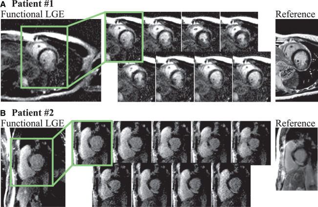
Late gadolinium enhancement (LGE) with cardiac magnetic resonance (CMR) imaging is the clinical reference for assessment of myocardial scar and focal fibrosis. However, current LGE techniques are confined to imaging of a single cardiac phase, which hampers assessment of scar motility and does not allow cross-comparison between multiple phases. In this work, we investigate a three step approach to obtain cardiac phase-resolved LGE images: (1) Acquisition of cardiac phase-resolved imaging data with varying weighting. (2) Generation of semi-quantitative maps for each cardiac phase. (3) Synthetization of LGE contrast to obtain functional LGE images. The proposed method is evaluated in phantom imaging, six healthy subjects at 3T and 20 patients at 1.5T. Phantom imaging at 3T demonstrates consistent contrast throughout the cardiac cycle with a coefficient of variation of 2.55 ± 0.42%. results show reliable LGE contrast with thorough suppression of the myocardial tissue is healthy subjects. The contrast between blood and myocardium showed moderate variation throughout the cardiac cycle in healthy subjects (coefficient of variation 18.2 ± 3.51%). Images were acquired at 40-60 ms and 80 ms temporal resolution, at 3T and 1.5, respectively. Functional LGE images acquired in patients with myocardial scar visualized scar tissue throughout the cardiac cycle, albeit at noticeably lower imaging resolution and noise resilience than the reference technique. The proposed technique bears the promise of integrating the advantages of phase-resolved CMR with LGE imaging, but further improvements in the acquisition quality are warranted for clinical use.

摘要

心脏磁共振成像(CMR)的延迟钆增强(LGE)是评估心肌瘢痕和局灶性纤维化的临床参考标准。然而,目前的LGE技术仅限于对单个心动周期进行成像,这妨碍了对瘢痕运动性的评估,并且不允许在多个心动周期之间进行交叉比较。在这项研究中,我们研究了一种三步法来获取心动周期分辨的LGE图像:(1)采集具有不同加权的心动周期分辨成像数据。(2)为每个心动周期生成半定量图。(3)合成LGE对比剂以获得功能性LGE图像。该方法在体模成像、6名3T的健康受试者和20名1.5T的患者中进行了评估。3T的体模成像显示在整个心动周期中对比度一致,变异系数为2.55±0.42%。结果表明,在健康受试者中,LGE对比可靠,心肌组织得到充分抑制。在健康受试者中,血液与心肌之间的对比度在整个心动周期中呈现中等程度的变化(变异系数为18.2±3.51%)。分别在3T和1.5T下,以40 - 60毫秒和80毫秒的时间分辨率采集图像。在心肌瘢痕患者中获取的功能性LGE图像在整个心动周期中均能显示瘢痕组织,尽管其成像分辨率和噪声耐受性明显低于参考技术。所提出的技术有望整合心动周期分辨CMR与LGE成像的优势,但为了临床应用,仍需进一步提高采集质量。

https://cdn.ncbi.nlm.nih.gov/pmc/blobs/f136/9557076/5cc4345a3ea2/fcvm-09-917180-g0008.jpg
https://cdn.ncbi.nlm.nih.gov/pmc/blobs/f136/9557076/692a6e82f459/fcvm-09-917180-g0001.jpg
https://cdn.ncbi.nlm.nih.gov/pmc/blobs/f136/9557076/128e6a042202/fcvm-09-917180-g0002.jpg
https://cdn.ncbi.nlm.nih.gov/pmc/blobs/f136/9557076/2c13c0bdc601/fcvm-09-917180-g0003.jpg
https://cdn.ncbi.nlm.nih.gov/pmc/blobs/f136/9557076/8f3f3d189ecc/fcvm-09-917180-g0004.jpg
https://cdn.ncbi.nlm.nih.gov/pmc/blobs/f136/9557076/6aa863c793b5/fcvm-09-917180-g0005.jpg
https://cdn.ncbi.nlm.nih.gov/pmc/blobs/f136/9557076/853f93e48adf/fcvm-09-917180-g0006.jpg
https://cdn.ncbi.nlm.nih.gov/pmc/blobs/f136/9557076/087e9042ad8f/fcvm-09-917180-g0007.jpg
https://cdn.ncbi.nlm.nih.gov/pmc/blobs/f136/9557076/5cc4345a3ea2/fcvm-09-917180-g0008.jpg

相似文献

1
Cardiac phase-resolved late gadolinium enhancement imaging.心脏相位分辨延迟钆增强成像。
Front Cardiovasc Med. 2022 Sep 29;9:917180. doi: 10.3389/fcvm.2022.917180. eCollection 2022.
2
Functional LGE Imaging: Cardiac Phase-Resolved Assessment of Focal Fibrosis.功能磁共振延迟强化成像:局灶性纤维化的心脏相位分辨评估
Annu Int Conf IEEE Eng Med Biol Soc. 2019 Jul;2019:3999-4003. doi: 10.1109/EMBC.2019.8857759.
3
Free-breathing combined three-dimensional phase sensitive late gadolinium enhancement and T1 mapping for myocardial tissue characterization.自由呼吸联合三维相位敏感晚期钆增强和T1映射用于心肌组织特征分析。
Magn Reson Med. 2015 Oct;74(4):1032-41. doi: 10.1002/mrm.25495. Epub 2014 Oct 16.
4
Synthetic late gadolinium enhancement cardiac magnetic resonance for diagnosing myocardial scar.用于诊断心肌瘢痕的合成延迟钆增强心脏磁共振成像
Scand Cardiovasc J. 2018 Jun;52(3):127-132. doi: 10.1080/14017431.2018.1449960. Epub 2018 Mar 16.
5
Comprehensive evaluation of macroscopic and microscopic myocardial fibrosis by cardiac MR: intra-individual comparison of gadobutrol versus gadoterate meglumine.心脏磁共振对心肌宏观和微观纤维化的综合评估:钆布醇与钆特酸葡胺的个体内比较。
Eur Radiol. 2019 Aug;29(8):4357-4367. doi: 10.1007/s00330-018-5956-3. Epub 2019 Jan 7.
6
Quantitative assessment of myocardial fibrosis in an age-related rat model by ex vivo late gadolinium enhancement magnetic resonance imaging with histopathological correlation.通过离体钆增强磁共振成像及组织病理学相关性对年龄相关大鼠模型中心肌纤维化进行定量评估。
Comput Biol Med. 2015 Oct 1;65:103-13. doi: 10.1016/j.compbiomed.2015.07.027. Epub 2015 Aug 8.
7
Free-Breathing Motion-Corrected Single-Shot Phase-Sensitive Inversion Recovery Late-Gadolinium-Enhancement Imaging: A Prospective Study of Image Quality in Patients with Hypertrophic Cardiomyopathy.自由呼吸运动校正单次激发相位敏感反转恢复晚期钆增强成像:肥厚型心肌病患者图像质量的前瞻性研究。
Korean J Radiol. 2021 Jul;22(7):1044-1053. doi: 10.3348/kjr.2020.1296. Epub 2021 Apr 1.
8
Feasibility study of a single breath-hold, 3D mDIXON pulse sequence for late gadolinium enhancement imaging of ischemic scar.单屏气 3D mDIXON 脉冲序列用于缺血性瘢痕晚期钆增强成像的可行性研究。
J Magn Reson Imaging. 2019 May;49(5):1437-1445. doi: 10.1002/jmri.26519. Epub 2018 Dec 31.
9
T(1) mapping for the diagnosis of acute myocarditis using CMR: comparison to T2-weighted and late gadolinium enhanced imaging.CMR 用于诊断急性心肌炎的 T(1) 映射:与 T2 加权和晚期钆增强成像的比较。
JACC Cardiovasc Imaging. 2013 Oct;6(10):1048-1058. doi: 10.1016/j.jcmg.2013.03.008. Epub 2013 Sep 4.
10
Quantitative T1 Mapping for Detecting Microvascular Obstruction in Reperfused Acute Myocardial Infarction: Comparison with Late Gadolinium Enhancement Imaging.定量 T1 映射检测再灌注急性心肌梗死中的微血管阻塞:与晚期钆增强成像的比较。
Korean J Radiol. 2020 Aug;21(8):978-986. doi: 10.3348/kjr.2019.0736.

引用本文的文献

1
Recent Progress of Cardiac MRI for Nuclear Medicine Professionals.核医学专业人员的心脏磁共振成像最新进展
Nucl Med Mol Imaging. 2024 Dec;58(7):431-448. doi: 10.1007/s13139-024-00850-9. Epub 2024 Feb 14.
2
Motion-compensated T mapping in cardiovascular magnetic resonance imaging: a technical review.心血管磁共振成像中的运动补偿T映射:技术综述
Front Cardiovasc Med. 2023 Sep 8;10:1160183. doi: 10.3389/fcvm.2023.1160183. eCollection 2023.

本文引用的文献

1
Free-breathing simultaneous , , and quantification in the myocardium.心肌自由呼吸状态下的同步[具体项目1]、[具体项目2]和[具体项目3]定量分析。 (注:原文中“ , , and ”部分内容缺失,以上为补充完整后的意译)
Magn Reson Med. 2021 Sep;86(3):1226-1240. doi: 10.1002/mrm.28753. Epub 2021 Mar 29.
2
Cardiac cine magnetic resonance fingerprinting for combined ejection fraction, T and T quantification.心脏电影磁共振指纹技术用于联合射血分数、T 值和 T* 值定量测量。
NMR Biomed. 2020 Aug;33(8):e4323. doi: 10.1002/nbm.4323. Epub 2020 Jun 5.
3
Simultaneous multi-slice T1 mapping using MOLLI with blipped CAIPIRINHA bSSFP.
使用带扰相的 CAIPIRINHA bSSFP 的 MOLLI 进行的同时多层 T1 映射。
Magn Reson Imaging. 2023 Jan;95:90-102. doi: 10.1016/j.mri.2020.03.006. Epub 2020 Apr 15.
4
Low-Rank Tensor Models for Improved Multi-Dimensional MRI: Application to Dynamic Cardiac Mapping.用于改进多维磁共振成像的低秩张量模型:在动态心脏映射中的应用
IEEE Trans Comput Imaging. 2019;6:194-207. doi: 10.1109/tci.2019.2940916. Epub 2019 Sep 12.
5
Deep Learning for Cardiac Image Segmentation: A Review.用于心脏图像分割的深度学习:综述
Front Cardiovasc Med. 2020 Mar 5;7:25. doi: 10.3389/fcvm.2020.00025. eCollection 2020.
6
Standardized cardiovascular magnetic resonance imaging (CMR) protocols: 2020 update.标准化心血管磁共振成像(CMR)协议:2020 年更新。
J Cardiovasc Magn Reson. 2020 Feb 24;22(1):17. doi: 10.1186/s12968-020-00607-1.
7
Sliding window reduced FOV reconstruction for real-time cardiac imaging.滑动窗口减小实时心脏成像的视场重建。
Z Med Phys. 2020 Aug;30(3):236-244. doi: 10.1016/j.zemedi.2020.01.001. Epub 2020 Feb 14.
8
Free-running cardiac magnetic resonance fingerprinting: Joint T1/T2 map and Cine imaging.自由运行心脏磁共振指纹技术:T1/T2 联合图谱和电影成像。
Magn Reson Imaging. 2020 May;68:173-182. doi: 10.1016/j.mri.2020.02.005. Epub 2020 Feb 13.
9
FULLY AUTOMATIC SEGMENTATION OF THE RIGHT VENTRICLE VIA MULTI-TASK DEEP NEURAL NETWORKS.通过多任务深度神经网络实现右心室的全自动分割
Proc IEEE Int Conf Acoust Speech Signal Process. 2018 Apr;2018:6677-6681. doi: 10.1109/ICASSP.2018.8461556. Epub 2018 Sep 13.
10
Reconstruction techniques for cardiac cine MRI.心脏电影磁共振成像的重建技术。
Insights Imaging. 2019 Sep 23;10(1):100. doi: 10.1186/s13244-019-0754-2.