D' Oria Salvatore, Giraldi David, Flores Daniel Andres Alvarado, Murrone Domenico, D' Angelo Vincenzo, Chaurasia Bipin
Department of Neurosurgery, Neurosurgical Unit of Miulli Hospital, Acquaviva Delle Fonti, Italy.
Department of Neurosurgery, Neurosurgical Unit of Azienda Ospedaliero Universitaria Consorziale Policlinico Di Bari, Italy.
J Craniovertebr Junction Spine. 2022 Jul-Sep;13(3):265-270. doi: 10.4103/jcvjs.jcvjs_87_22. Epub 2022 Sep 14.
Hemangioblastomas (HBs) are rare lesions accounting for 1%-5% of all spinal cord tumors, and are mostly associated with Von Hippel-Lindau (VHL) syndrome. Localization in the cauda equina is uncommon.
In this manuscript, we aimed to describe a rare case of sporadic intradural extramedullary HB of the cauda equina and present a literature review.
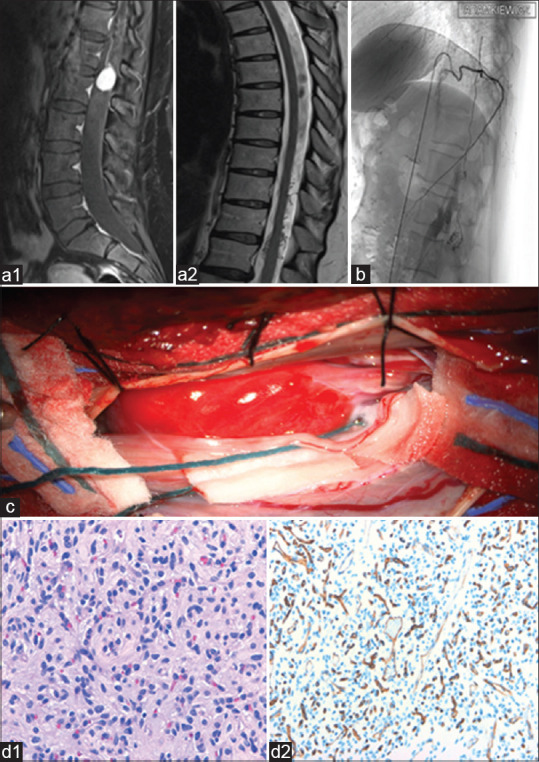
A systematic research was performed on PubMed, MEDLINE, and Google Scholar, using the keywords "spinal HB" and "cauda equina tumors." The previous literature is integrated by the description of the present case. A 49-year-old female presented in August 2020 to our institution with a magnetic resonance imaging (MRI) which showed an intradural mass at L1/2 level and angiography that showing a nidus of serpiginous vessels inside the lesion. Symptoms were right sciatica and paresthesia in right L5 radicular dermatome for more than 3 months. Neurological examination revealed claudicatio spinalis and hypoesthesia on right L5 dermatome and weakness of right anterior tibialis muscle. Microsurgical en bloc resection of lesion was performed with adjuvant neurophysiological intraoperative monitoring. The histological examination provided the diagnosis of HB.
After surgery, symptoms and neurological impairment gradually improved. Postoperative MRI showed no residual tumor.
Although intradural extramedullary HB of the cauda equina without VHL syndrome is a rare pathological entity, this diagnosis must be taken in consideration when a mass affects cauda equina. Preoperative embolization is an option to minimize intraoperative bleeding. Radiosurgery seems to prevent recurrences when the tumor is not completely excised. A complete surgical removal of the lesion is usually possible and it leads to a low likelihood of recurrence.
血管母细胞瘤(HBs)是罕见病变,占所有脊髓肿瘤的1%-5%,且大多与冯·希佩尔-林道(VHL)综合征相关。位于马尾的情况并不常见。
在本手稿中,我们旨在描述一例罕见的散发性马尾硬膜内髓外HB病例,并进行文献综述。
在PubMed、MEDLINE和谷歌学术上进行了系统研究,使用关键词“脊髓HB”和“马尾肿瘤”。通过对本病例的描述整合先前的文献。一名49岁女性于2020年8月就诊于我院,磁共振成像(MRI)显示L1/2水平硬膜内肿块,血管造影显示病变内有蜿蜒血管团。症状为右侧坐骨神经痛和右侧L5神经根皮节感觉异常超过3个月。神经学检查显示脊髓间歇性跛行、右侧L5皮节感觉减退以及右侧胫前肌无力。在辅助神经生理术中监测下对病变进行了显微外科整块切除。组织学检查确诊为HB。
术后症状和神经功能障碍逐渐改善。术后MRI显示无残留肿瘤。
尽管无VHL综合征的马尾硬膜内髓外HB是一种罕见的病理实体,但当肿块累及马尾时必须考虑这一诊断。术前栓塞是减少术中出血的一种选择。当肿瘤未完全切除时,放射外科似乎可预防复发。通常能够完整手术切除病变,且复发可能性低。