Department of Internal Medicine, Kyungpook National University Hospital, Daegu, Korea.
School of Medicine, Kyungpook National University, Daegu, Korea.
J Clin Hypertens (Greenwich). 2022 Nov;24(11):1451-1460. doi: 10.1111/jch.14583. Epub 2022 Oct 21.
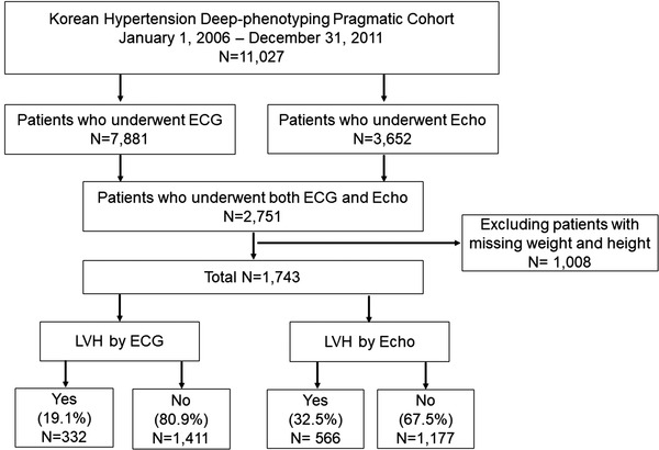
In patients with hypertension, left ventricular hypertrophy (LVH) represents a risk factor for cardiovascular disease and asymptomatic organ damage. Currently, electrocardiography (ECG) and two-dimensional echocardiography (Echo) are the most widely used methods for LVH evaluation. This study aimed to compare the long-term outcomes of LVH, as evaluated by ECG and Echo, in patients with hypertension. Patients diagnosed with hypertension as a primary disease between 2006 and 2011 were enrolled in the Korean Hypertension Cohort study. The study finally included 1743 patients who underwent both ECG and Echo. The primary endpoint was defined as the composite of major adverse cardiovascular events (MACEs) or death. Overall, LVH was identified in 747 patients. The patients were categorized into four groups according to the detection of LVH by ECG or Echo: No LVH (n = 996), LVH diagnosed by ECG alone (n = 181), LVH diagnosed by Echo alone (n = 415), LVH diagnosed by both ECG and Echo (n = 151). After adjusting for variables, the incidence of MACEs or death was significantly greater in patients with LVH diagnosed by ECG alone (hazards ratio [HR]: 1.69; 95% confidence interval [CI]: 1.22-2.35; P = .001), LVH diagnosed by Echo alone (HR: 1.54; 95% CI: 1.16-2.05; P = .002), and LVH diagnosed by both ECG and Echo (HR: 1.87; 95% CI: 1.18-2.94; P = .002) than in those with no LVH. Both ECG and Echo are efficient diagnostic tools for LVH and useful for long-term risk stratification. Additional Echo evaluation for LVH is helpful for predicting long-term outcomes only in patients without LVH diagnosis by ECG.
在高血压患者中,左心室肥厚(LVH)是心血管疾病和无症状器官损伤的危险因素。目前,心电图(ECG)和二维超声心动图(Echo)是评估 LVH 的最广泛使用的方法。本研究旨在比较心电图和超声心动图评估高血压患者 LVH 的长期结果。2006 年至 2011 年间被诊断为原发性高血压的患者被纳入韩国高血压队列研究。研究最终纳入了 1743 名同时接受心电图和超声心动图检查的患者。主要终点定义为主要不良心血管事件(MACEs)或死亡的复合终点。总体而言,747 名患者存在 LVH。根据心电图或超声心动图检测到的 LVH,患者被分为四组:无 LVH(n=996)、仅心电图诊断的 LVH(n=181)、仅超声心动图诊断的 LVH(n=415)、心电图和超声心动图均诊断的 LVH(n=151)。在校正了变量后,仅心电图诊断的 LVH(危险比[HR]:1.69;95%置信区间[CI]:1.22-2.35;P=0.001)、仅超声心动图诊断的 LVH(HR:1.54;95%CI:1.16-2.05;P=0.002)和心电图和超声心动图均诊断的 LVH(HR:1.87;95%CI:1.18-2.94;P=0.002)患者的 MACEs 或死亡发生率显著高于无 LVH 患者。心电图和超声心动图都是 LVH 的有效诊断工具,有助于进行长期风险分层。对于无心电图诊断 LVH 的患者,额外的超声心动图评估 LVH 有助于预测长期预后。