Mother Infant Research Institute (MIRI), Tufts Medical Center, Boston, MA, USA.
Department of Pediatrics, Tufts Medical Center, Boston, MA, USA.
Pediatr Res. 2023 Feb;93(3):604-611. doi: 10.1038/s41390-022-02357-5. Epub 2022 Oct 24.
Preclinical data demonstrate that opioids modulate brain reward signaling through an inflammatory cascade, but this relationship has yet to be studied in opioid-exposed neonates.
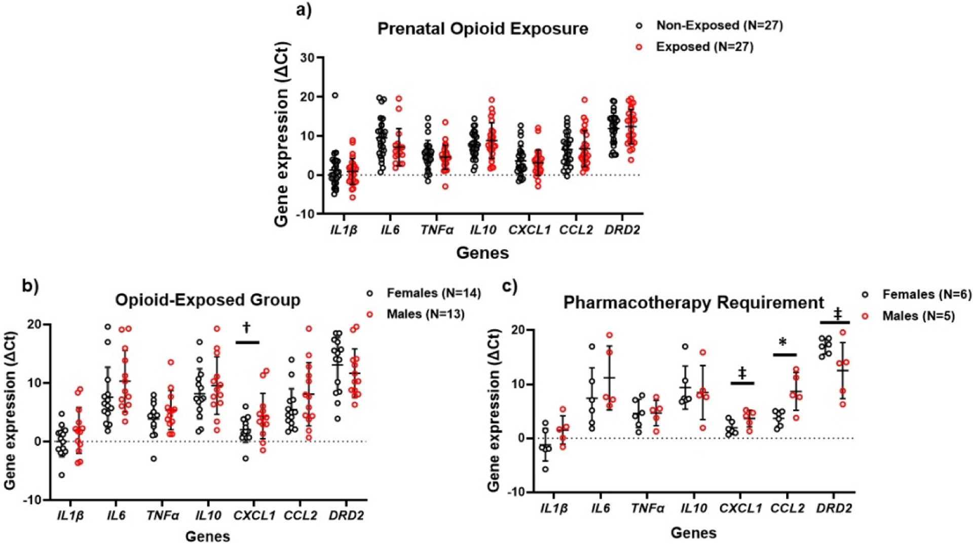
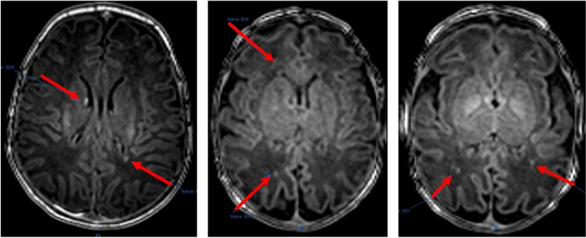
Saliva samples of 54 opioid-exposed and sex- and age-matched non-exposed neonates underwent transcriptomic analysis of inflammatory and reward genes. A subset of 22 neonates underwent brain magnetic resonance imaging (MRI) to evaluate white matter injury commonly associated with inflammatory response. Gene expression and brain MRI were compared between opioid- and non-exposed neonates and further stratified by sex and pharmacotherapy need.
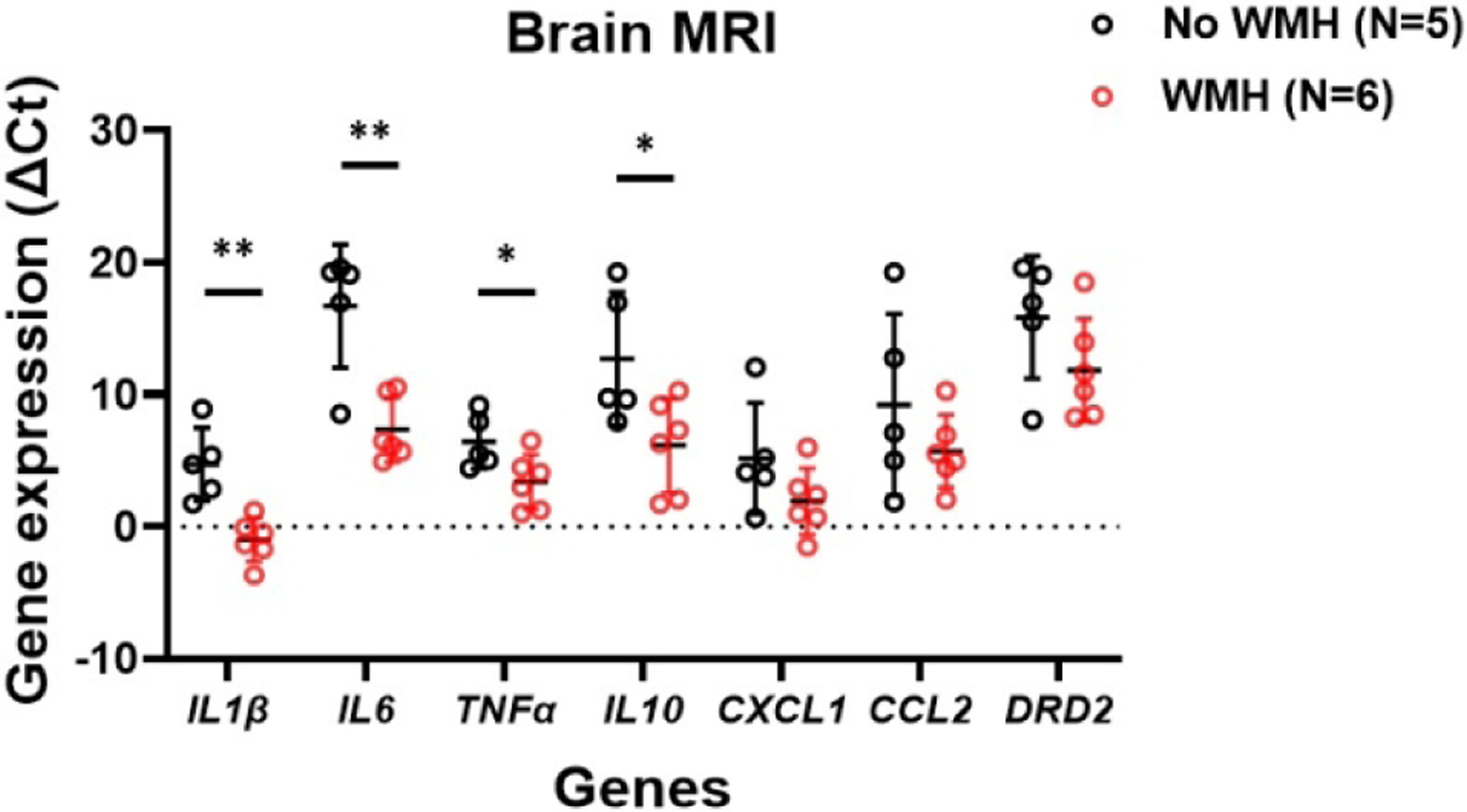
Opioid-exposed females regardless of pharmacotherapy need had higher expression of inflammatory genes than their male counterparts, with notable differences in the expression of CCL2 and CXCL1 in females requiring pharmacotherapy (p = 0.01 and 0.06, respectively). Opioid-exposed males requiring pharmacotherapy had higher expression of DRD2 than exposed females (p = 0.07), validating our prior research. Higher expression of IL1β, IL6, TNFα, and IL10 was seen in opioid-exposed neonates with T1 white matter hyperintensity (WMH) compared to exposed neonates without WMH (p < 0.05).
Prenatal opioid exposure may promote inflammation resulting in changes in reward signaling and white matter injury in the developing brain, with unique sex-specific effects. The actions of opioids through non-neuronal pathways need further investigation.
Opioid-exposed neonates are at risk for punctate T1 white matter hyperintensity (WMH). Females carry a greater propensity for WMH. Salivary transcriptomic data showed significantly higher expression of inflammatory genes in opioid-exposed neonates with WMH than those without WMH, irrespective of pharmacotherapy need. Adding to prior studies, our findings suggest that prenatal opioid exposure may modulate white matter injury and reward signaling through a pro-inflammatory process that is sex specific. This novel study highlights the short-term molecular and structural effects of prenatal opioids and the need to elucidate the long-term impact of prenatal opioid exposure.
临床前数据表明,阿片类药物通过炎症级联反应调节大脑奖励信号,但这一关系尚未在暴露于阿片类药物的新生儿中进行研究。
对 54 名暴露于阿片类药物的新生儿和性别及年龄匹配的非暴露于阿片类药物的新生儿的唾液样本进行了炎症和奖励基因的转录组分析。对 22 名新生儿进行了脑磁共振成像 (MRI) 检查,以评估与炎症反应相关的常见白质损伤。比较了阿片类药物暴露和非暴露新生儿之间的基因表达和脑 MRI,并根据性别和药物治疗需求进一步分层。
无论药物治疗需求如何,暴露于阿片类药物的女性的炎症基因表达均高于男性,尤其是需要药物治疗的女性的 CCL2 和 CXCL1 表达差异显著(分别为 p=0.01 和 0.06)。需要药物治疗的暴露于阿片类药物的男性的 DRD2 表达高于暴露于阿片类药物的女性(p=0.07),这验证了我们之前的研究。与没有白质高信号(WMH)的暴露于阿片类药物的新生儿相比,有 T1 白质高信号(WMH)的暴露于阿片类药物的新生儿的 IL1β、IL6、TNFα 和 IL10 表达更高(p<0.05)。
产前阿片类药物暴露可能会促进炎症,导致发育中大脑的奖励信号和白质损伤发生变化,具有独特的性别特异性影响。阿片类药物通过非神经元途径的作用需要进一步研究。
暴露于阿片类药物的新生儿有发生点状 T1 白质高信号(WMH)的风险。女性更容易发生 WMH。唾液转录组数据显示,无论是否需要药物治疗,有 WMH 的暴露于阿片类药物的新生儿的炎症基因表达明显高于无 WMH 的新生儿。除了之前的研究,我们的研究结果表明,产前阿片类药物暴露可能通过炎症过程来调节白质损伤和奖励信号,这种炎症过程具有性别特异性。这项新研究强调了产前阿片类药物的短期分子和结构影响,需要阐明产前阿片类药物暴露的长期影响。