• 文献检索
  • 文档翻译
  • 深度研究
  • 学术资讯
  • Suppr Zotero 插件Zotero 插件
  • 邀请有礼
  • 套餐&价格
  • 历史记录
应用&插件
Suppr Zotero 插件Zotero 插件浏览器插件Mac 客户端Windows 客户端微信小程序
定价
高级版会员购买积分包购买API积分包
服务
文献检索文档翻译深度研究API 文档MCP 服务
关于我们
关于 Suppr公司介绍联系我们用户协议隐私条款
关注我们

Suppr 超能文献

核心技术专利:CN118964589B侵权必究
粤ICP备2023148730 号-1Suppr @ 2026

文献检索

告别复杂PubMed语法,用中文像聊天一样搜索,搜遍4000万医学文献。AI智能推荐,让科研检索更轻松。

立即免费搜索

文件翻译

保留排版,准确专业,支持PDF/Word/PPT等文件格式,支持 12+语言互译。

免费翻译文档

深度研究

AI帮你快速写综述,25分钟生成高质量综述,智能提取关键信息,辅助科研写作。

立即免费体验

利用深度学习技术在视盘环扫描中提前检测到视神经炎。

Prior optic neuritis detection on peripapillary ring scans using deep learning.

机构信息

Experimental and Clinical Research Center, a cooperation between the Max Delbrück Center for Molecular Medicine in the Helmholtz Association and Charité Universitätsmedizin Berlin, corporate member of Freie Universität Berlin and Humboldt-Universität zu Berlin, Berlin, Germany.

Nocturne GmbH, Berlin, Germany.

出版信息

Ann Clin Transl Neurol. 2022 Nov;9(11):1682-1691. doi: 10.1002/acn3.51632. Epub 2022 Oct 25.

DOI:10.1002/acn3.51632
PMID:36285339
原文链接:https://pmc.ncbi.nlm.nih.gov/articles/PMC9639624/
Abstract

BACKGROUND

The diagnosis of multiple sclerosis (MS) requires demyelinating events that are disseminated in time and space. Peripapillary retinal nerve fiber layer (pRNFL) thickness as measured by optical coherence tomography (OCT) distinguishes eyes with a prior history of acute optic neuritis (ON) and may provide evidence to support a demyelinating attack.

OBJECTIVE

To investigate whether a deep learning (DL)-based network can distinguish between eyes with prior ON and healthy control (HC) eyes using peripapillary ring scans.

METHODS

We included 1033 OCT scans from 415 healthy eyes (213 HC subjects) and 510 peripapillary ring scans from 164 eyes with prior acute ON (140 patients with MS). Data were split into 70% training, 15% validation, and 15% test data. We included 102 OCT scans from 80 healthy eyes (40 HC) and 61 scans from 40 ON eyes (31 MS patients) from an independent second center. Receiver operating characteristic curve analyses with area under the curve (AUC) were used to investigate performance.

RESULTS

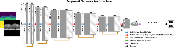
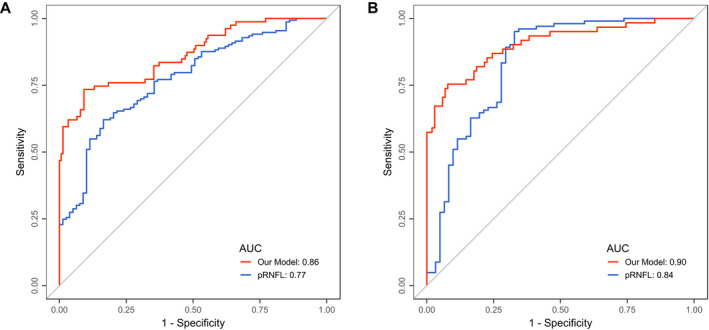
We used a dilated residual convolutional neural network for the classification. The final network had an accuracy of 0.85 and an AUC of 0.86, whereas pRNFL only had an AUC of 0.77 in recognizing ON eyes. Using data from a second center, the network achieved an accuracy of 0.77 and an AUC of 0.90 compared to pRNFL, which had an AUC of 0.84.

INTERPRETATION

DL-based disease classification of prior ON is feasible and has the potential to outperform thickness-based classification of eyes with and without history of prior ON.

摘要

背景

多发性硬化症(MS)的诊断需要时间和空间上弥散的脱髓鞘事件。光学相干断层扫描(OCT)测量的视盘周围视网膜神经纤维层(pRNFL)厚度可区分有急性视神经炎(ON)病史的眼睛,并可为脱髓鞘发作提供证据。

目的

研究基于深度学习(DL)的网络是否可以使用视盘周围环扫来区分有既往 ON 病史的眼睛和健康对照(HC)眼睛。

方法

我们纳入了来自 415 只健康眼(213 例 HC 受试者)的 1033 个 OCT 扫描和来自 164 只既往急性 ON 眼(140 例 MS 患者)的 510 个视盘周围环扫。数据分为 70%训练、15%验证和 15%测试数据。我们纳入了来自第二个中心的 80 只健康眼(40 例 HC)的 102 个 OCT 扫描和来自 40 只 ON 眼(31 例 MS 患者)的 61 个扫描。使用曲线下面积(AUC)的接收者操作特征曲线分析来研究性能。

结果

我们使用扩张残差卷积神经网络进行分类。最终网络的准确率为 0.85,AUC 为 0.86,而 pRNFL 在识别 ON 眼时的 AUC 仅为 0.77。使用第二个中心的数据,网络的准确率为 0.77,AUC 为 0.90,而 pRNFL 的 AUC 为 0.84。

解释

基于 DL 的既往 ON 疾病分类是可行的,并且有可能优于有无既往 ON 病史的眼睛的基于厚度的分类。

https://cdn.ncbi.nlm.nih.gov/pmc/blobs/bdcc/9639624/48891e7ee0ea/ACN3-9-1682-g003.jpg
https://cdn.ncbi.nlm.nih.gov/pmc/blobs/bdcc/9639624/bc885e459d47/ACN3-9-1682-g004.jpg
https://cdn.ncbi.nlm.nih.gov/pmc/blobs/bdcc/9639624/da84fba025e2/ACN3-9-1682-g002.jpg
https://cdn.ncbi.nlm.nih.gov/pmc/blobs/bdcc/9639624/136388de20f4/ACN3-9-1682-g001.jpg
https://cdn.ncbi.nlm.nih.gov/pmc/blobs/bdcc/9639624/48891e7ee0ea/ACN3-9-1682-g003.jpg

相似文献

1
Prior optic neuritis detection on peripapillary ring scans using deep learning.利用深度学习技术在视盘环扫描中提前检测到视神经炎。
Ann Clin Transl Neurol. 2022 Nov;9(11):1682-1691. doi: 10.1002/acn3.51632. Epub 2022 Oct 25.
2
OCT retinal nerve fiber layer thickness differentiates acute optic neuritis from MOG antibody-associated disease and Multiple Sclerosis: RNFL thickening in acute optic neuritis from MOGAD vs MS.光学相干断层扫描(OCT)测量的视网膜神经纤维层厚度可区分急性视神经炎与MOG抗体相关疾病及多发性硬化症:MOG抗体相关疾病与多发性硬化症所致急性视神经炎的视网膜神经纤维层增厚情况比较
Mult Scler Relat Disord. 2022 Feb;58:103525. doi: 10.1016/j.msard.2022.103525. Epub 2022 Jan 11.
3
A multi-regression approach to improve optical coherence tomography diagnostic accuracy in multiple sclerosis patients without previous optic neuritis.一种多回归方法,用于提高没有先前视神经炎的多发性硬化症患者的光学相干断层扫描诊断准确性。
Neuroimage Clin. 2022;34:103010. doi: 10.1016/j.nicl.2022.103010. Epub 2022 Apr 16.
4
Optical Coherence Tomography for the Detection of Remote Optic Neuritis in Multiple Sclerosis.光学相干断层扫描用于检测多发性硬化症中的远隔性视神经炎
J Neuroimaging. 2016 May;26(3):283-8. doi: 10.1111/jon.12326. Epub 2016 Jan 15.
5
Use of retinal optical coherence tomography to differentiate suspected neuromyelitis optica spectrum disorder from multiple sclerosis: A cross-sectional study.利用视网膜光学相干断层扫描鉴别疑似视神经脊髓炎谱系障碍与多发性硬化症:一项横断面研究。
Mult Scler Relat Disord. 2022 Dec;68:104160. doi: 10.1016/j.msard.2022.104160. Epub 2022 Sep 7.
6
Peripapillary retinal nerve fiber layer thickness measured by optical coherence tomography in different clinical subtypes of multiple sclerosis.光学相干断层扫描测量多发性硬化不同临床亚型的视盘周围视网膜神经纤维层厚度。
Mult Scler Relat Disord. 2019 Jan;27:260-268. doi: 10.1016/j.msard.2018.11.003. Epub 2018 Nov 5.
7
The Role of Optical Coherence Tomography Criteria and Machine Learning in Multiple Sclerosis and Optic Neuritis Diagnosis.光学相干断层扫描标准和机器学习在多发性硬化症和视神经炎诊断中的作用。
Neurology. 2022 Sep 13;99(11):e1100-e1112. doi: 10.1212/WNL.0000000000200883. Epub 2022 Jun 28.
8
Retinal imaging by laser polarimetry and optical coherence tomography evidence of axonal degeneration in multiple sclerosis.通过激光偏振imetry和光学相干断层扫描进行视网膜成像:多发性硬化症中轴突退变的证据
Arch Neurol. 2008 Jul;65(7):924-8. doi: 10.1001/archneur.65.7.924.
9
Optical coherence tomography angiography enhances the detection of optic nerve damage in multiple sclerosis.光学相干断层扫描血管造影增强多发性硬化症视神经损伤的检测。
Br J Ophthalmol. 2018 Apr;102(4):520-524. doi: 10.1136/bjophthalmol-2017-310477. Epub 2017 Aug 16.
10
Retinal layer segmentation in multiple sclerosis: a systematic review and meta-analysis.多发性硬化症中的视网膜层分割:系统评价和荟萃分析。
Lancet Neurol. 2017 Oct;16(10):797-812. doi: 10.1016/S1474-4422(17)30278-8. Epub 2017 Sep 12.

引用本文的文献

1
Effectiveness of oral prednisone tapering following intravenous methylprednisolone for acute optic neuritis in multiple sclerosis.口服泼尼松龙逐渐减量治疗多发性硬化急性视神经炎静脉注射甲泼尼龙的疗效。
PLoS One. 2023 Dec 7;18(12):e0288366. doi: 10.1371/journal.pone.0288366. eCollection 2023.

本文引用的文献

1
The Role of Optical Coherence Tomography Criteria and Machine Learning in Multiple Sclerosis and Optic Neuritis Diagnosis.光学相干断层扫描标准和机器学习在多发性硬化症和视神经炎诊断中的作用。
Neurology. 2022 Sep 13;99(11):e1100-e1112. doi: 10.1212/WNL.0000000000200883. Epub 2022 Jun 28.
2
Intraretinal Layer Segmentation Using Cascaded Compressed U-Nets.使用级联压缩U型网络进行视网膜内层分割
J Imaging. 2022 May 17;8(5):139. doi: 10.3390/jimaging8050139.
3
Modular deep neural networks for automatic quality control of retinal optical coherence tomography scans.
用于视网膜光学相干断层扫描自动质量控制的模块化深度神经网络。
Comput Biol Med. 2022 Feb;141:104822. doi: 10.1016/j.compbiomed.2021.104822. Epub 2021 Sep 20.
4
Long-term outcomes in patients presenting with optic neuritis: Analyses of the MSBase registry.视神经炎患者的长期预后:MSBase 注册研究分析。
J Neurol Sci. 2021 Nov 15;430:118067. doi: 10.1016/j.jns.2021.118067. Epub 2021 Sep 3.
5
Artificial intelligence extension of the OSCAR-IB criteria.OSCAR-IB 标准的人工智能扩展。
Ann Clin Transl Neurol. 2021 Jul;8(7):1528-1542. doi: 10.1002/acn3.51320. Epub 2021 May 19.
6
APOSTEL 2.0 Recommendations for Reporting Quantitative Optical Coherence Tomography Studies.APOSTEL 2.0 关于报告定量光学相干断层扫描研究的建议。
Neurology. 2021 Jul 13;97(2):68-79. doi: 10.1212/WNL.0000000000012125. Epub 2021 Apr 28.
7
Optic Nerve Topography in Multiple Sclerosis Diagnosis: The Utility of Visual Evoked Potentials.多发性硬化症诊断中的视神经地形图:视觉诱发电位的应用。
Neurology. 2021 Jan 26;96(4):e482-e490. doi: 10.1212/WNL.0000000000011339. Epub 2020 Dec 16.
8
Optic Nerve in Multiple Sclerosis Diagnostic Criteria: An Aye to the Eyes?多发性硬化诊断标准中的视神经:眼睛的关键指标?
Neurology. 2021 Jan 26;96(4):139-140. doi: 10.1212/WNL.0000000000011344. Epub 2020 Dec 16.
9
Retinal asymmetry in multiple sclerosis.多发性硬化症中的视网膜不对称性。
Brain. 2021 Feb 12;144(1):224-235. doi: 10.1093/brain/awaa361.
10
Optical coherence tomography for detection of asymptomatic optic nerve lesions in clinically isolated syndrome.光学相干断层扫描在临床孤立综合征无症状性视神经病变中的应用
Neurology. 2020 Aug 11;95(6):e733-e744. doi: 10.1212/WNL.0000000000009832. Epub 2020 Jul 28.