Medical Physics & Biomedical Engineering Department, School of Medicine, Tehran University of Medical Sciences (TUMS), Tehran, Iran.
Medical Imaging Center, Motahari Hospital, Jahrom University of Medical Sciences (JUMS), Jahrom, Iran.
Contrast Media Mol Imaging. 2022 Oct 12;2022:5425851. doi: 10.1155/2022/5425851. eCollection 2022.
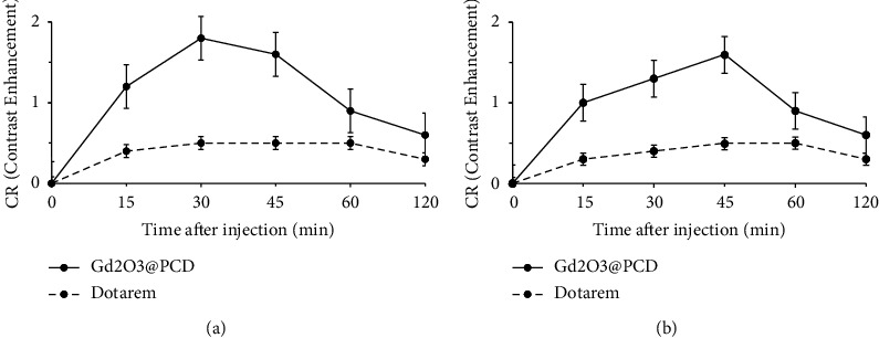
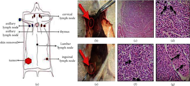
Axillary lymph node detection is crucial to staging and prognosis of the lymph node metastatic spread in breast cancer. Currently, lymphoscintigraphy and blue dye, as the conventional methods to localize sentinel lymph nodes (SLNs), are invasive and can only be performed during surgery. This study has had a novel hybrid gadolinium oxide nanoparticle coating with Cyclodextrin-based polyester as a high-relaxivity T magnetic resonance molecular imaging (MRMI) contrast agent (CA). Twelve female BALB/c mice were randomly divided into three groups of four mice; each group was injected with 4T cells to obtain metastasis lymph nodes and diagnosed by using the 3D TW (VIBE) MRI (Siemens 3T, Prisma). The synthesized GdO@PCD nanoparticles with a suitable particle size range of 20-40 nm have had much higher longitudinal relaxivity ( ) for GdO@PCD and Gd-DOTA (Dotarem) with the values of 3.98 mM·s ± 0.003 and 2.71 mM·s ± 0.005, respectively. Identical MR images in coronal views were subsequently obtained to create time-intensity curves of the right axillary lymph nodes and to measure the contrast ratio (CR). The peak CR and qualitative assessment of axillary lymph nodes at five-time points were evaluated. After subcutaneous injection, the contrast ratio of axillary lymph node and tumor in mice exhibited CR peak of GdO@PCD and Dotarem with the values of 2.21 ± 0.06 and 0.40 ± 0.004 for lymph node and 2.54 ± 0.04 and 1.21 ± 0.007 for the tumor, respectively. Furthermore, the lumbar-aortic lymph node is weakly visible in the original coronal image. In conclusion, the use of GdO@PCD nanoparticles as novel MRMI CAs enables high resolution for the detection of lymph node metastasis in mice with the potential capability for breast cancer diagnostic imaging.
腋窝淋巴结检测对于乳腺癌的淋巴结转移分期和预后至关重要。目前,淋巴闪烁显像和蓝染作为定位前哨淋巴结 (SLN) 的常规方法具有侵袭性,只能在手术过程中进行。本研究采用了一种新型的混合氧化钆纳米粒子涂层,其表面涂有基于环糊精的聚酯,作为一种高弛豫率 T 磁共振分子成像 (MRMI) 造影剂 (CA)。将 12 只雌性 BALB/c 小鼠随机分为三组,每组 4 只小鼠;每组均注射 4T 细胞以获得转移淋巴结,并使用 3D TW (VIBE) MRI(西门子 3T,Prisma)进行诊断。合成的 GdO@PCD 纳米粒子具有合适的粒径范围(20-40nm),其纵向弛豫率()显著高于 GdO@PCD 和 Gd-DOTA(Dotarem),分别为 3.98mM·s±0.003 和 2.71mM·s±0.005。随后获得冠状位相同的 MR 图像,以创建右腋窝淋巴结的时间-强度曲线,并测量对比比(CR)。评估了五个时间点的峰值 CR 和腋窝淋巴结的定性评估。皮下注射后,小鼠腋窝淋巴结和肿瘤的对比比显示 GdO@PCD 和 Dotarem 的 CR 峰值分别为 2.21±0.06 和 0.40±0.004(淋巴结)和 2.54±0.04 和 1.21±0.007(肿瘤)。此外,在原始冠状图像中,腰椎-主动脉淋巴结也可见。总之,GdO@PCD 纳米粒子作为新型的 MRMI CA,可实现对小鼠淋巴结转移的高分辨率检测,具有乳腺癌诊断成像的潜力。