Department of Diagnostic and Interventional Radiology, Medical Faculty, University Dusseldorf, Moorenstrasse 5, 40225, Dusseldorf, Germany.
Department of Gynecology and Obstetrics, University Hospital Essen, University of Duisburg-Essen, 45147, Essen, Germany.
Eur J Nucl Med Mol Imaging. 2022 Feb;49(3):992-1001. doi: 10.1007/s00259-021-05502-0. Epub 2021 Sep 3.
To compare CT, MRI, and [F]-fluorodeoxyglucose positron emission tomography ([F]-FDG PET/MRI) for nodal status, regarding quantity and location of metastatic locoregional lymph nodes in patients with newly diagnosed breast cancer.
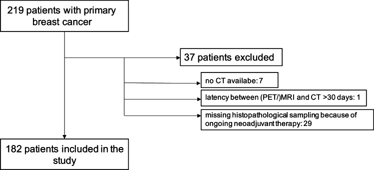
One hundred eighty-two patients (mean age 52.7 ± 11.9 years) were included in this prospective double-center study. Patients underwent dedicated contrast-enhanced chest/abdomen/pelvis computed tomography (CT) and whole-body ([F]-FDG PET/) magnet resonance imaging (MRI). Thoracal datasets were evaluated separately regarding quantity, lymph node station (axillary levels I-III, supraclavicular, internal mammary chain), and lesion character (benign vs. malign). Histopathology served as reference standard for patient-based analysis. Patient-based and lesion-based analyses were compared by a McNemar test. Sensitivity, specificity, positive and negative predictive values, and accuracy were assessed for all three imaging modalities.
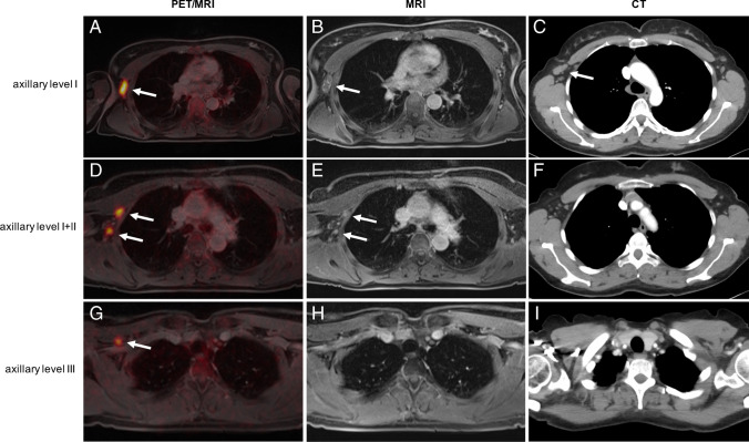
On a patient-based analysis, PET/MRI correctly detected significantly more nodal positive patients than MRI (p < 0.0001) and CT (p < 0.0001). No statistically significant difference was seen between CT and MRI. PET/MRI detected 193 lesions in 75 patients (41.2%), while MRI detected 123 lesions in 56 patients (30.8%) and CT detected 104 lesions in 50 patients, respectively. Differences were statistically significant on a lesion-based analysis (PET/MRI vs. MRI, p < 0.0001; PET/MRI vs. CT, p < 0.0001; MRI vs. CT, p = 0.015). Subgroup analysis for different lymph node stations showed that PET/MRI detected significantly more lymph node metastases than MRI and CT in each location (axillary levels I-III, supraclavicular, mammary internal chain). MRI was superior to CT only in axillary level I (p = 0.0291).
[F]-FDG PET/MRI outperforms CT or MRI in detecting nodal involvement on a patient-based analysis and on a lesion-based analysis. Furthermore, PET/MRI was superior to CT or MRI in detecting lymph node metastases in all lymph node stations. Of all the tested imaging modalities, PET/MRI showed the highest sensitivity, whereas CT showed the lowest sensitivity, but was most specific.
比较 CT、MRI 和 [F]-氟脱氧葡萄糖正电子发射断层扫描 ([F]-FDG PET/MRI) 对新诊断乳腺癌患者局部区域淋巴结转移的淋巴结状态,包括转移淋巴结的数量和位置。
本前瞻性双中心研究纳入 182 例患者(平均年龄 52.7±11.9 岁)。患者接受了专用对比增强胸部/腹部/骨盆 CT(CT)和全身 ([F]-FDG PET/) 磁共振成像(MRI)检查。胸部数据集分别评估淋巴结数量、淋巴结站(腋窝 I-III 水平、锁骨上、内乳链)和病变特征(良性与恶性)。患者层面分析以组织病理学为参考标准。采用 McNemar 检验比较患者层面和病变层面分析。评估了三种成像方式的敏感性、特异性、阳性预测值、阴性预测值和准确性。
在患者层面分析中,PET/MRI 比 MRI(p<0.0001)和 CT(p<0.0001)更准确地检测到更多的淋巴结阳性患者。CT 和 MRI 之间无统计学差异。PET/MRI 在 75 例患者中检测到 193 个病变(41.2%),MRI 在 56 例患者中检测到 123 个病变(30.8%),CT 在 50 例患者中检测到 104 个病变。在病变层面分析中,差异具有统计学意义(PET/MRI 与 MRI,p<0.0001;PET/MRI 与 CT,p<0.0001;MRI 与 CT,p=0.015)。不同淋巴结站的亚组分析显示,PET/MRI 在每个部位(腋窝 I-III 水平、锁骨上、内乳链)比 MRI 和 CT 更准确地检测到淋巴结转移。MRI 仅在腋窝 I 水平优于 CT(p=0.0291)。
在患者层面分析和病变层面分析中,[F]-FDG PET/MRI 比 CT 或 MRI 更准确地检测淋巴结受累情况。此外,PET/MRI 在所有淋巴结站均优于 CT 或 MRI 检测淋巴结转移。在所有测试的成像方式中,PET/MRI 显示出最高的敏感性,而 CT 显示出最低的敏感性,但具有最高的特异性。