van der Zee Sygrid, Kanel Prabesh, Müller Martijn L T M, van Laar Teus, Bohnen Nicolaas I
Department of Radiology, University of Michigan, Ann Arbor, MI, United States.
Department of Neurology, University of Groningen, University Medical Center Groningen, Groningen, Netherlands.
Front Aging Neurosci. 2022 Oct 20;14:1006567. doi: 10.3389/fnagi.2022.1006567. eCollection 2022.
Degeneration of the cholinergic system plays an important role in cognitive impairment in Parkinson's disease (PD). Positron emission tomography (PET) imaging using the presynaptic vesicular acetylcholine transporter (VAChT) tracer [F]Fluoroethoxybenzovesamicol ([F]FEOBV) allows for regional assessment of cholinergic innervation. The purpose of this study was to perform a data-driven analysis to identify co-varying cholinergic regions and to evaluate the relationship of these with cognitive functioning in PD.
A total of 87 non-demented PD patients (77% male, mean age 67.9 ± 7.6 years, disease duration 5.8 ± 4.6 years) and 27 healthy control (HC) subjects underwent [F]FEOBV brain PET imaging and neuropsychological assessment. A volume-of-interest based factor analysis was performed for both groups to identify cholinergic principal components (PCs).
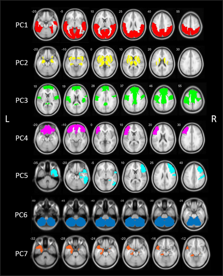
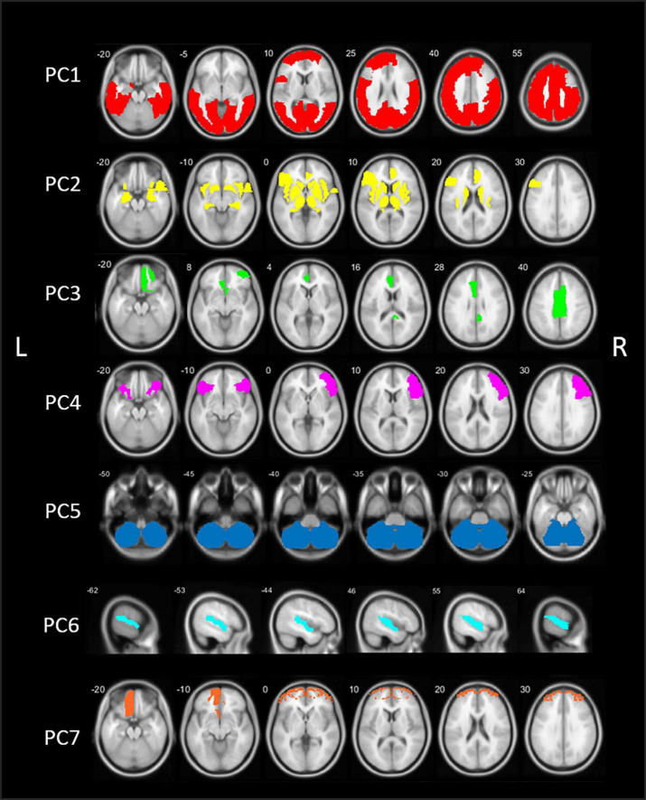
Seven main PCs were identified for the PD group: (1) bilateral posterior cortex, (2) bilateral subcortical, (3) bilateral centro-cingulate, (4) bilateral frontal, (5) right-sided fronto-temporal, (6) cerebellum, and (7) predominantly left sided temporal regions. A complementary principal component analysis (PCA) analysis in the control group showed substantially different cholinergic covarying patterns. A multivariate linear regression analyses demonstrated PC3, PC5, and PC7, together with motor impairment score, as significant predictors for cognitive functioning in PD. PC3 showed most robust correlations with cognitive functioning ( < 0.001).
A data-driven approach identified covarying regions in the bilateral peri-central and cingulum cortex as a key determinant of cognitive impairment in PD. Cholinergic vulnerability of the centro-cingulate network appears to be disease-specific for PD rather than being age-related. The cholinergic system may be an important contributor to regional and large scale neural networks involved in cognitive functioning.
胆碱能系统的退化在帕金森病(PD)的认知障碍中起重要作用。使用突触前囊泡乙酰胆碱转运体(VAChT)示踪剂[F]氟乙氧基苯并维司那明([F]FEOBV)的正电子发射断层扫描(PET)成像可对胆碱能神经支配进行区域评估。本研究的目的是进行数据驱动分析,以识别共同变化的胆碱能区域,并评估这些区域与PD患者认知功能的关系。
共有87例非痴呆PD患者(男性占77%,平均年龄67.9±7.6岁,病程5.8±4.6年)和27名健康对照(HC)受试者接受了[F]FEOBV脑PET成像和神经心理学评估。对两组进行基于感兴趣区的因子分析,以识别胆碱能主成分(PCs)。
PD组确定了7个主要PCs:(1)双侧后皮质,(2)双侧皮质下,(3)双侧中央扣带回,(4)双侧额叶,(5)右侧额颞叶,(6)小脑,(7)主要为左侧颞叶区域。对照组的补充主成分分析(PCA)显示出明显不同的胆碱能共同变化模式。多变量线性回归分析表明,PC3、PC5和PC7以及运动障碍评分是PD患者认知功能的重要预测指标。PC3与认知功能的相关性最强(<0.001)。
数据驱动方法确定双侧中央周围和扣带回皮质中共同变化的区域是PD认知障碍的关键决定因素。中央扣带回网络的胆碱能易损性似乎是PD特有的,而非与年龄相关。胆碱能系统可能是参与认知功能的区域和大规模神经网络的重要贡献者。