Meah Mohammed N, Bularga Anda, Tzolos Evangelos, Chapman Andrew R, Daghem Marwa, Hung John D, Chiong Justin, Taggart Caelan, Wereski Ryan, Gray Alasdair, Dweck Marc R, Roobottom Carl, Curzen Nick, Kardos Attila, Felmeden Dirk, Mills Nicholas L, Slomka Piotr J, Newby David E, Dey Damini, Williams Michelle C
British Heart Foundation Centre of Cardiovascular Science, University of Edinburgh, Edinburgh, Scotland (M.N.M., A.B., E.T., A.R.C., M.D., J.D.H., J.C., C.T., R.W., A.G., M.R.D., N.L.M., D.E.N., M.C.W.); Usher Institute, University of Edinburgh, Edinburgh, Scotland (A.G., N.L.M.); University Hospital Plymouth, Plymouth, England (C.R.); Faculty of Medicine, University of Southampton, Southampton, England (N.C.); University Hospital Southampton, Southampton, England (N.C.); Department of Cardiology, Milton Keynes University Hospital, School of Sciences and Medicine, University of Buckingham, Buckingham, England (A.K.); Torbay and South Devon NHS Foundation Trust, Torquay, England (D.F.); Departments of Medicine and Biomedical Sciences, Cedars-Sinai Medical Center, Los Angeles, Calif (P.J.S., D.D.); and Edinburgh Imaging, Queen's Medical Research Institute University of Edinburgh, Edinburgh, Scotland (D.E.N., M.C.W.).
Radiol Cardiothorac Imaging. 2022 Oct 27;4(5):e220081. doi: 10.1148/ryct.220081. eCollection 2022 Oct.
To determine whether quantitative plaque characterization by using CT coronary angiography (CTCA) can discriminate between type 1 and type 2 myocardial infarction.
This was a secondary analysis of two prospective studies (ClinicalTrials.gov registration nos. NCT03338504 [2014-2019] and NCT02284191 [2018-2020]) that performed blinded quantitative plaque analysis on findings from CTCA in participants with type 1 myocardial infarction, type 2 myocardial infarction, and chest pain without myocardial infarction. Logistic regression analyses were performed to identify predictors of type 1 myocardial infarction.
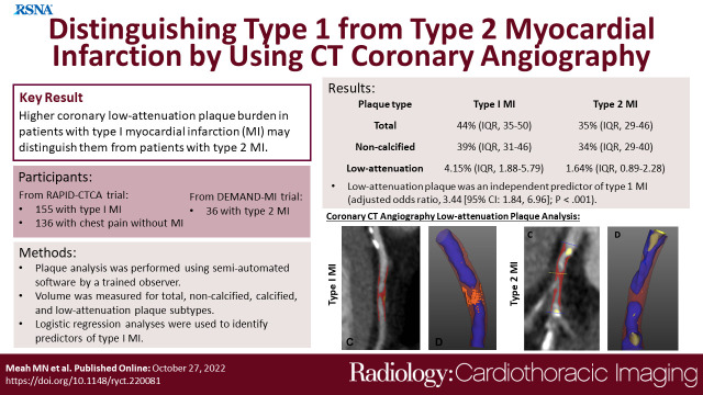
Overall, 155 participants (mean age, 64 years ± 12 [SD]; 114 men) and 36 participants (mean age, 67 years ± 12; 19 men) had type 1 and type 2 myocardial infarction, respectively, and 136 participants (62 years ± 12; 78 men) had chest pain without myocardial infarction. Participants with type 1 myocardial infarction had greater total (median, 44% [IQR: 35%-50%] vs 35% [IQR: 29%-46%]), noncalcified (39% [IQR: 31%-46%] vs 34% [IQR: 29%-40%]), and low-attenuation (4.15% [IQR: 1.88%-5.79%] vs 1.64% [IQR: 0.89%-2.28%]) plaque burdens ( < .05 for all) than those with type 2. Participants with type 2 myocardial infarction had similar low-attenuation plaque burden to those with chest pain without myocardial infarction (P = .4). Low-attenuation plaque was an independent predictor of type 1 myocardial infarction (adjusted odds ratio, 3.44 [95% CI: 1.84, 6.96]; < .001), with better discrimination than noncalcified plaque burden and maximal area of coronary stenosis (C statistic, 0.75 [95% CI: 0.67, 0.83] vs 0.62 [95% CI: 0.53, 0.71] and 0.61 [95% CI: 0.51, 0.70] respectively; ≤ .001 for both).
Higher low-attenuation coronary plaque burden in patients with type 1 myocardial infarction may help distinguish these patients from those with type 2 myocardial infarction. Ischemia/Infarction, CT Angiography, Quantitative CTClinical trial registration nos. NCT03338504 and NCT02284191 © RSNA, 2022.
确定使用CT冠状动脉造影(CTCA)进行定量斑块特征分析是否能够区分1型和2型心肌梗死。
这是对两项前瞻性研究(ClinicalTrials.gov注册号分别为NCT03338504[2014 - 2019]和NCT02284191[2018 - 2020])的二次分析,对1型心肌梗死、2型心肌梗死和无心肌梗死的胸痛患者的CTCA结果进行了盲法定量斑块分析。进行逻辑回归分析以确定1型心肌梗死的预测因素。
总体而言,分别有155名参与者(平均年龄64岁±12[标准差];114名男性)和36名参与者(平均年龄67岁±12;19名男性)患有1型和2型心肌梗死,136名参与者(62岁±12;78名男性)有胸痛但无心肌梗死。1型心肌梗死患者的总斑块(中位数,44%[四分位间距:35% - 50%]对35%[四分位间距:29% - 46%])、非钙化斑块(39%[四分位间距:31% - 46%]对34%[四分位间距:29% - 40%])和低衰减斑块(4.15%[四分位间距:1.88% - 5.79%]对1.64%[四分位间距:0.89% - 2.28%])负担均高于2型患者(所有P均<0.05)。2型心肌梗死患者的低衰减斑块负担与无心肌梗死的胸痛患者相似(P = 0.4)。低衰减斑块是1型心肌梗死的独立预测因素(调整后的优势比,3.44[95%置信区间:1.84,6.96];P<0.001),其鉴别能力优于非钙化斑块负担和冠状动脉狭窄最大面积(C统计量分别为0.75[95%置信区间:0.67,0.83]对0.62[95%置信区间:0.53,0.71]和0.61[95%置信区间:0.51,0.70];两者P均≤0.001)。
1型心肌梗死患者较高的低衰减冠状动脉斑块负担可能有助于将这些患者与2型心肌梗死患者区分开来。缺血/梗死、CT血管造影、定量CT临床试验注册号NCT03338504和NCT02284191 ©RSNA,2022