Wijesinghe Harshima Disvini, Wijesinghe Gayani Kokila, Fernando Deepika, de Silva Chandu
Department of Pathology, Faculty of Medicine, University of Colombo, Colombo, Sri Lanka.
Department of Parasitology, Faculty of Medicine, University of Kelaniya, Sri Lanka.
Clin Pathol. 2022 Nov 2;15:2632010X221134804. doi: 10.1177/2632010X221134804. eCollection 2022 Jan-Dec.
is the causative organism of leishmaniasis in Sri Lanka. Studies on the immunopathology of leishmaniasis due to L. donovani are limited. The objective of this study was to describe the immunopathological characteristics of cutaneous leishmaniasis in a cohort of Sri Lankan patients.
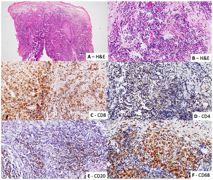
Fifty skin biopsies of cutaneous leishmaniasis confirmed by detection of organisms by histology, culture, slit-skin smear, and/or polymerase chain reaction were reviewed. The inflammatory infiltrate was characterized by immunohistochemical staining for CD4, CD8, CD20, and CD68. Associations and correlations between immunohistochemical staining pattern and the parasitic load, and patterns of inflammation were determined.
The majority of biopsies showed a CD8+/CD4- T lymphocyte predominant infiltrate (84%, n = 42). A CD68 predominant infiltrate was seen in 16%(n = 8). The mean percentage of CD8+, CD4+, CD20+, and CD68+ inflammatory cells in the biopsies were 56.1% (SD = 16.5%), 2.6% (SD = 4.5%), 12.3% (SD = 10.9%), and 25.7% (SD = 15.8%) respectively. There was no association between the predominant inflammatory cell and the degree of inflammation ( = .173), presence of high RPI ( = .922), MRI( = .367) or presence of granuloma ( = .247).The percentage of CD4+ cells showed a positive correlation with granuloma formation (Correlation coefficient = .411, = .03). The percentage of CD20+ cells in the infiltrate showed a positive correlation with the degree of inflammation (Correlation coefficient = .491, = .02) and the RPI (Correlation coefficient = .334, = .018).
Skin biopsies from cutaneous leishmaniasis due to infection showed a CD8+/CD4- predominant infiltrate. This is similar to the findings of studies on cutaneous leishmaniasis due to some other species and suggests that the cytotoxic T cell response plays a role in infections due to .
杜氏利什曼原虫是斯里兰卡利什曼病的病原体。关于杜氏利什曼原虫所致利什曼病免疫病理学的研究有限。本研究的目的是描述一组斯里兰卡皮肤利什曼病患者的免疫病理特征。
回顾了50例经组织学、培养、皮肤涂片和/或聚合酶链反应检测到病原体而确诊的皮肤利什曼病皮肤活检标本。通过对CD4、CD8、CD20和CD68进行免疫组织化学染色来表征炎症浸润。确定免疫组织化学染色模式与寄生虫负荷及炎症模式之间的关联和相关性。
大多数活检标本显示以CD8+/CD4-T淋巴细胞为主的浸润(84%,n = 42)。16%(n = 8)的标本可见以CD68为主的浸润。活检标本中CD8+、CD4+、CD20+和CD68+炎症细胞的平均百分比分别为56.1%(标准差 = 16.5%)、2.6%(标准差 = 4.5%)、12.3%(标准差 = 10.9%)和25.7%(标准差 = 15.8%)。主要炎症细胞与炎症程度(P = 0.173)、高寄生虫感染率指数(P = 0.922)、磁共振成像(P = 0.367)或肉芽肿的存在(P = 0.247)之间无关联。CD4+细胞百分比与肉芽肿形成呈正相关(相关系数 = 0.411,P = 0.03)。浸润中CD20+细胞百分比与炎症程度呈正相关(相关系数 = 0.491,P = 0.02),与寄生虫感染率指数呈正相关(相关系数 = 0.334,P = 0.018)。
杜氏利什曼原虫感染所致皮肤利什曼病的皮肤活检显示以CD8+/CD4-为主的浸润。这与其他一些物种所致皮肤利什曼病的研究结果相似,表明细胞毒性T细胞反应在杜氏利什曼原虫感染中起作用。