• 文献检索
  • 文档翻译
  • 深度研究
  • 学术资讯
  • Suppr Zotero 插件Zotero 插件
  • 邀请有礼
  • 套餐&价格
  • 历史记录
应用&插件
Suppr Zotero 插件Zotero 插件浏览器插件Mac 客户端Windows 客户端微信小程序
定价
高级版会员购买积分包购买API积分包
服务
文献检索文档翻译深度研究API 文档MCP 服务
关于我们
关于 Suppr公司介绍联系我们用户协议隐私条款
关注我们

Suppr 超能文献

核心技术专利:CN118964589B侵权必究
粤ICP备2023148730 号-1Suppr @ 2026

文献检索

告别复杂PubMed语法,用中文像聊天一样搜索,搜遍4000万医学文献。AI智能推荐,让科研检索更轻松。

立即免费搜索

文件翻译

保留排版,准确专业,支持PDF/Word/PPT等文件格式,支持 12+语言互译。

免费翻译文档

深度研究

AI帮你快速写综述,25分钟生成高质量综述,智能提取关键信息,辅助科研写作。

立即免费体验

弥漫性皮肤利什曼病患者的 CD8 细胞表现出功能衰竭:后者可被 TLR2 激动剂体外逆转。

CD8 cells of patients with diffuse cutaneous leishmaniasis display functional exhaustion: the latter is reversed, in vitro, by TLR2 agonists.

机构信息

Departamento de Medicina Experimental, Facultad de Medicina, Universidad Nacional Autónoma de México, Hospital General de México OD, México Distrito Federal, Mexico.

出版信息

PLoS Negl Trop Dis. 2010 Nov 2;4(11):e871. doi: 10.1371/journal.pntd.0000871.

DOI:10.1371/journal.pntd.0000871
PMID:21072232
原文链接:https://pmc.ncbi.nlm.nih.gov/articles/PMC2970528/
Abstract

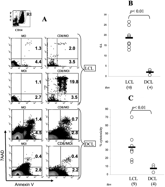
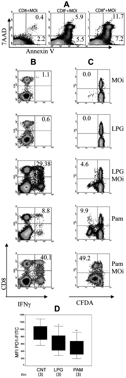
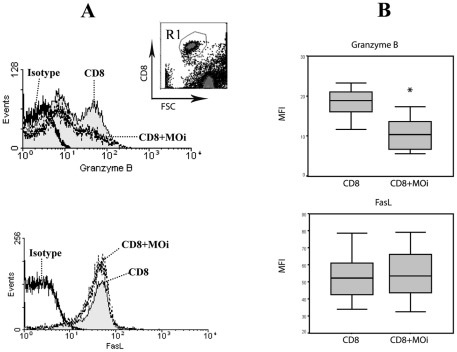
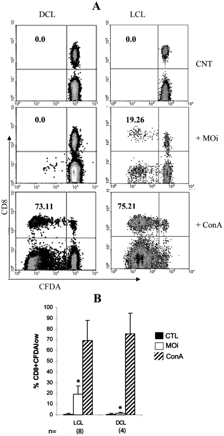
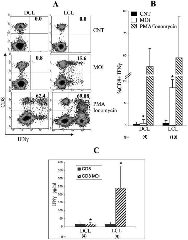
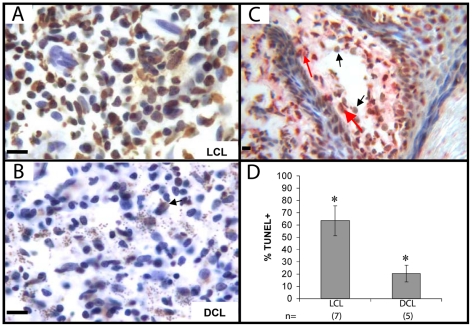
Leishmania mexicana (Lm) causes localized (LCL) and diffuse (DCL) cutaneous leishmaniasis. DCL patients have a poor cellular immune response leading to chronicity. It has been proposed that CD8 T lymphocytes (CD8) play a crucial role in infection clearance, although the role of CD8 cytotoxicity in disease control has not been elucidated. Lesions of DCL patients have been shown to harbor low numbers of CD8, as compared to patients with LCL, and leishmanicidal treatment restores CD8 numbers. The marked response of CD8 towards Leishmania parasites led us to analyze possible functional differences between CD8 from patients with LCL and DCL. We compared IFNγ production, antigen-specific proliferation, and cytotoxicity of CD8 purified from PBMC against autologous macrophages (MO) infected with Leishmania mexicana (MOi). Additionally, we analyzed tissue biopsies from both groups of patients for evidence of cytotoxicity associated with apoptotic cells in the lesions. We found that CD8 cell of DCL patients exhibited low cytotoxicity, low antigen-specific proliferation and low IFNγ production when stimulated with MOi, as compared to LCL patients. Additionally, DCL patients had significantly less TUNEL+ cells in their lesions. These characteristics are similar to cellular "exhaustion" described in chronic infections. We intended to restore the functional capacity of CD8 cells of DCL patients by preincubating them with TLR2 agonists: Lm lipophosphoglycan (LPG) or Pam3Cys. Cytotoxicity against MOi, antigen-specific proliferation and IFNγ production were restored with both stimuli, whereas PD-1 (a molecule associated with cellular exhaustion) expression, was reduced. Our work suggests that CD8 response is associated with control of Lm infection in LCL patients and that chronic infection in DCL patients leads to a state of CD8 functional exhaustion, which could facilitate disease spread. This is the first report that shows the presence of functionally exhausted CD8 T lymphocytes in DCL patients and, additionally, that pre-stimulation with TLR2 ligands can restore the effector mechanisms of CD8 T lymphocytes from DCL patients against Leishmania mexicana-infected macrophages.

摘要

墨西哥利什曼原虫(Lm)可引起局限性(LCL)和弥漫性(DCL)皮肤利什曼病。DCL 患者的细胞免疫反应较差,导致疾病慢性化。有人提出,CD8 T 淋巴细胞(CD8)在清除感染中发挥关键作用,尽管 CD8 细胞毒性在疾病控制中的作用尚未阐明。与 LCL 患者相比,DCL 患者的病变中 CD8 数量较低,而利什曼病治疗可恢复 CD8 数量。CD8 对利什曼原虫的显著反应促使我们分析 LCL 和 DCL 患者的 CD8 之间可能存在的功能差异。我们比较了从 PBMC 中纯化的 CD8 针对感染了墨西哥利什曼原虫的自体巨噬细胞(MOi)产生 IFNγ、抗原特异性增殖和细胞毒性的能力。此外,我们还分析了两组患者的组织活检,以寻找病变中与凋亡细胞相关的细胞毒性的证据。我们发现,与 LCL 患者相比,DCL 患者的 CD8 细胞在受到 MOi 刺激时表现出低细胞毒性、低抗原特异性增殖和低 IFNγ产生。此外,DCL 患者病变中的 TUNEL+细胞明显较少。这些特征类似于慢性感染中描述的细胞“衰竭”。我们试图通过用 TLR2 激动剂预先孵育 DCL 患者的 CD8 细胞来恢复其功能能力:Lm 脂磷壁酸(LPG)或 Pam3Cys。两种刺激物均可恢复对 MOi 的细胞毒性、抗原特异性增殖和 IFNγ 产生,而 PD-1(与细胞衰竭相关的分子)表达降低。我们的工作表明,CD8 反应与 LCL 患者中 Lm 感染的控制有关,而 DCL 患者中的慢性感染导致 CD8 功能衰竭状态,这可能促进疾病传播。这是首次报道表明 DCL 患者存在功能衰竭的 CD8 T 淋巴细胞,并且,预先刺激 TLR2 配体可以恢复 DCL 患者针对感染了墨西哥利什曼原虫的巨噬细胞的 CD8 T 淋巴细胞的效应机制。

https://cdn.ncbi.nlm.nih.gov/pmc/blobs/5f57/2970528/bea157a8331c/pntd.0000871.g006.jpg
https://cdn.ncbi.nlm.nih.gov/pmc/blobs/5f57/2970528/430630c3178d/pntd.0000871.g001.jpg
https://cdn.ncbi.nlm.nih.gov/pmc/blobs/5f57/2970528/f7ef1ab2289c/pntd.0000871.g002.jpg
https://cdn.ncbi.nlm.nih.gov/pmc/blobs/5f57/2970528/db12915d0dce/pntd.0000871.g003.jpg
https://cdn.ncbi.nlm.nih.gov/pmc/blobs/5f57/2970528/5f28572e0384/pntd.0000871.g004.jpg
https://cdn.ncbi.nlm.nih.gov/pmc/blobs/5f57/2970528/3d65945e8bc0/pntd.0000871.g005.jpg
https://cdn.ncbi.nlm.nih.gov/pmc/blobs/5f57/2970528/bea157a8331c/pntd.0000871.g006.jpg

相似文献

1
CD8 cells of patients with diffuse cutaneous leishmaniasis display functional exhaustion: the latter is reversed, in vitro, by TLR2 agonists.弥漫性皮肤利什曼病患者的 CD8 细胞表现出功能衰竭:后者可被 TLR2 激动剂体外逆转。
PLoS Negl Trop Dis. 2010 Nov 2;4(11):e871. doi: 10.1371/journal.pntd.0000871.
2
NK cell activity differs between patients with localized and diffuse cutaneous leishmaniasis infected with Leishmania mexicana: a comparative study of TLRs and cytokines.感染墨西哥利什曼原虫的局限性和弥漫性皮肤利什曼病患者的自然杀伤细胞活性存在差异:Toll样受体和细胞因子的比较研究
PLoS One. 2014 Nov 14;9(11):e112410. doi: 10.1371/journal.pone.0112410. eCollection 2014.
3
Monocyte cytokine and costimulatory molecule expression in patients infected with Leishmania mexicana.感染墨西哥利什曼原虫患者的单核细胞细胞因子及共刺激分子表达
Parasite Immunol. 2007 Mar;29(3):117-26. doi: 10.1111/j.1365-3024.2006.00924.x.
4
Differential Regulation of l-Arginine Metabolism through Arginase 1 during Infection with Leishmania mexicana Isolates Obtained from Patients with Localized and Diffuse Cutaneous Leishmaniasis.通过 Arg1ase1 对 l-精氨酸代谢的差异调控在来源于局限性和皮肤弥散性利什曼病患者的 Leishmania mexicana 分离株感染中的作用。
Infect Immun. 2020 Jun 22;88(7). doi: 10.1128/IAI.00963-19.
5
Toll-like receptor 2 (TLR2) plays a role in controlling cutaneous leishmaniasis in vivo, but does not require activation by parasite lipophosphoglycan.Toll样受体2(TLR2)在体内控制皮肤利什曼病中发挥作用,但不需要被寄生虫脂磷壁酸激活。
Parasit Vectors. 2016 Oct 6;9(1):532. doi: 10.1186/s13071-016-1807-8.
6
Host and parasite responses in human diffuse cutaneous leishmaniasis caused by L. amazonensis.由 L. amazonensis 引起的人类弥漫性皮肤利什曼病中的宿主和寄生虫反应。
PLoS Negl Trop Dis. 2019 Mar 7;13(3):e0007152. doi: 10.1371/journal.pntd.0007152. eCollection 2019 Mar.
7
[Immunopathology of American tegumentary leishmaniasis].[美洲皮肤利什曼病的免疫病理学]
Acta Cient Venez. 1998;49(1):42-56.
8
PD-L1 May Mediate T-Cell Exhaustion in a Case of Early Diffuse Leishmaniasis Caused by (L.) .PD-L1 可能介导了由 (L.)引起的早期弥漫性利什曼病中的 T 细胞耗竭。
Front Immunol. 2018 May 11;9:1021. doi: 10.3389/fimmu.2018.01021. eCollection 2018.
9
Exposure of phosphatidylserine on Leishmania amazonensis isolates is associated with diffuse cutaneous leishmaniasis and parasite infectivity.巴西利什曼原虫分离株磷脂酰丝氨酸暴露与皮肤利什曼病的弥漫性和寄生虫感染力有关。
PLoS One. 2012;7(5):e36595. doi: 10.1371/journal.pone.0036595. Epub 2012 May 4.
10
Eosinophils of patients with localized and diffuse cutaneous leishmaniasis: Differential response to Leishmania mexicana, with insights into mechanisms of damage inflicted upon the parasites by eosinophils.局限性和播散性皮肤利什曼病患者的嗜酸性粒细胞:对墨西哥利什曼原虫的差异反应,深入了解嗜酸性粒细胞对寄生虫造成损害的机制。
PLoS One. 2024 Feb 15;19(2):e0296887. doi: 10.1371/journal.pone.0296887. eCollection 2024.

引用本文的文献

1
Senescence-related genes are associated with the immunopathology signature of American tegumentary leishmaniasis lesions and may predict progression to mucosal leishmaniasis.衰老相关基因与美洲皮肤利什曼病病变的免疫病理学特征相关,并且可能预测向黏膜利什曼病的进展。
Clin Exp Immunol. 2025 Jan 21;219(1). doi: 10.1093/cei/uxae088.
2
CXCR5 and TIM-3 expressions define distinct exhausted T cell subsets in experimental cutaneous infection with .CXCR5 和 TIM-3 的表达在实验性皮肤感染 中定义了不同的耗竭性 T 细胞亚群。
Front Immunol. 2023 Aug 25;14:1231836. doi: 10.3389/fimmu.2023.1231836. eCollection 2023.
3
The Extraordinary Case of a Woman with a 30-Year-Long Diffuse Leishmaniasis Cured with One Single Ampoule of Intranasal Pentavalent Antimoniate.

本文引用的文献

1
TLR-9 signaling and TCR stimulation co-regulate CD8(+) T cell-associated PD-1 expression.TLR-9 信号和 TCR 刺激共同调节 CD8(+) T 细胞相关 PD-1 的表达。
Immunol Lett. 2009 Dec 2;127(1):60-7. doi: 10.1016/j.imlet.2009.09.002. Epub 2009 Sep 12.
2
The involvement of TLR2 in cytokine and reactive oxygen species (ROS) production by PBMCs in response to Leishmania major phosphoglycans (PGs).Toll样受体2(TLR2)参与单核细胞在响应硕大利什曼原虫磷酸化聚糖(PGs)时细胞因子和活性氧(ROS)的产生。
Parasitology. 2009 Sep;136(10):1193-9. doi: 10.1017/S0031182009990473. Epub 2009 Jul 27.
3
Leishmania infection inhibits cycloheximide-induced macrophage apoptosis in a strain-dependent manner.
一位患有长达30年弥漫性利什曼病的女性仅通过一剂鼻内注射五价锑酸盐就得以治愈的非凡病例。
Pathogens. 2023 Jun 29;12(7):890. doi: 10.3390/pathogens12070890.
4
Immunopathology of Cutaneous Leishmaniasis in a Cohort of Sri Lankan Patients.一组斯里兰卡患者皮肤利什曼病的免疫病理学
Clin Pathol. 2022 Nov 2;15:2632010X221134804. doi: 10.1177/2632010X221134804. eCollection 2022 Jan-Dec.
5
T Lymphocyte Exhaustion During Human and Experimental Visceral Leishmaniasis.人内脏利什曼病和实验性内脏利什曼病中的 T 淋巴细胞耗竭。
Front Immunol. 2022 May 2;13:835711. doi: 10.3389/fimmu.2022.835711. eCollection 2022.
6
Paradoxical immune response in leishmaniasis: The role of toll-like receptors in disease progression.利什曼病中的矛盾免疫反应: toll 样受体在疾病进展中的作用。
Parasite Immunol. 2022 Apr;44(4-5):e12910. doi: 10.1111/pim.12910. Epub 2022 Feb 28.
7
Successful treatment of diffuse cutaneous leishmaniasis caused by Leishmania amazonensis.成功治疗由亚马逊利什曼原虫引起的皮肤弥漫性利什曼病。
An Bras Dermatol. 2021 Sep-Oct;96(5):602-604. doi: 10.1016/j.abd.2021.03.003. Epub 2021 Jul 15.
8
Unraveling the Role of Immune Checkpoints in Leishmaniasis.解析免疫检查点在利什曼病中的作用。
Front Immunol. 2021 Mar 11;12:620144. doi: 10.3389/fimmu.2021.620144. eCollection 2021.
9
PD-1 Blockade Modulates Functional Activities of Exhausted-Like T Cell in Patients With Cutaneous Leishmaniasis.PD-1 阻断调节皮肤利什曼病患者衰竭样 T 细胞的功能活性。
Front Immunol. 2021 Mar 9;12:632667. doi: 10.3389/fimmu.2021.632667. eCollection 2021.
10
Programmed Cell Death Ligand (PD-L)-1 Contributes to the Regulation of CD4 T Effector and Regulatory T Cells in Cutaneous Leishmaniasis.程序性细胞死亡配体 1(PD-L)有助于调控皮肤利什曼病中的 CD4 效应 T 细胞和调节性 T 细胞。
Front Immunol. 2020 Oct 22;11:574491. doi: 10.3389/fimmu.2020.574491. eCollection 2020.
利什曼原虫感染以菌株依赖的方式抑制环己酰亚胺诱导的巨噬细胞凋亡。
Exp Parasitol. 2009 Sep;123(1):58-64. doi: 10.1016/j.exppara.2009.05.012. Epub 2009 Jun 3.
4
Chronic antigen stimulation alone is sufficient to drive CD8+ T cell exhaustion.仅慢性抗原刺激就足以驱动CD8 + T细胞耗竭。
J Immunol. 2009 Jun 1;182(11):6697-708. doi: 10.4049/jimmunol.0800997.
5
B7-H1 blockade increases survival of dysfunctional CD8(+) T cells and confers protection against Leishmania donovani infections.B7-H1阻断可提高功能失调的CD8(+) T细胞的存活率,并赋予对杜氏利什曼原虫感染的保护作用。
PLoS Pathog. 2009 May;5(5):e1000431. doi: 10.1371/journal.ppat.1000431. Epub 2009 May 15.
6
Cell death mechanisms induced by cytotoxic lymphocytes.细胞毒性淋巴细胞诱导的细胞死亡机制。
Cell Mol Immunol. 2009 Feb;6(1):15-25. doi: 10.1038/cmi.2009.3.
7
Leishmania mexicana: inhibition of camptothecin-induced apoptosis of monocyte-derived dendritic cells.墨西哥利什曼原虫:对喜树碱诱导的单核细胞衍生树突状细胞凋亡的抑制作用
Exp Parasitol. 2009 Mar;121(3):199-207. doi: 10.1016/j.exppara.2008.10.020. Epub 2008 Nov 18.
8
A critical role for direct TLR2-MyD88 signaling in CD8 T-cell clonal expansion and memory formation following vaccinia viral infection.在牛痘病毒感染后,TLR2-MyD88直接信号传导在CD8 T细胞克隆扩增和记忆形成中起关键作用。
Blood. 2009 Mar 5;113(10):2256-64. doi: 10.1182/blood-2008-03-148809. Epub 2008 Oct 23.
9
Functional restoration of HCV-specific CD8 T cells by PD-1 blockade is defined by PD-1 expression and compartmentalization.通过程序性死亡蛋白1(PD-1)阻断实现丙型肝炎病毒(HCV)特异性CD8 T细胞的功能恢复,这由PD-1表达和区室化所定义。
Gastroenterology. 2008 Jun;134(7):1927-37, 1937.e1-2. doi: 10.1053/j.gastro.2008.02.033. Epub 2008 Feb 17.
10
Toll-like receptor signalling on Tregs: to suppress or not to suppress?调节性T细胞上的Toll样受体信号传导:抑制还是不抑制?
Immunology. 2008 Aug;124(4):445-52. doi: 10.1111/j.1365-2567.2008.02871.x. Epub 2008 Jun 28.