Ricciardi Dario, Amitrano Federica, Coccia Armando, Todisco Vincenzo, Trojsi Francesca, Tedeschi Gioacchino, Cirillo Giovanni
I Division of Neurology and Neurophysiopathology, Department of Medical and Surgical Sciences, University of Campania "Luigi Vanvitelli", 80138 Naples, Italy.
Department of Information Technologies and Electrical Engineering, University of Naples "Federico II", 80125 Naples, Italy.
Brain Sci. 2022 Nov 7;12(11):1510. doi: 10.3390/brainsci12111510.
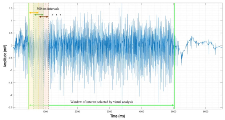
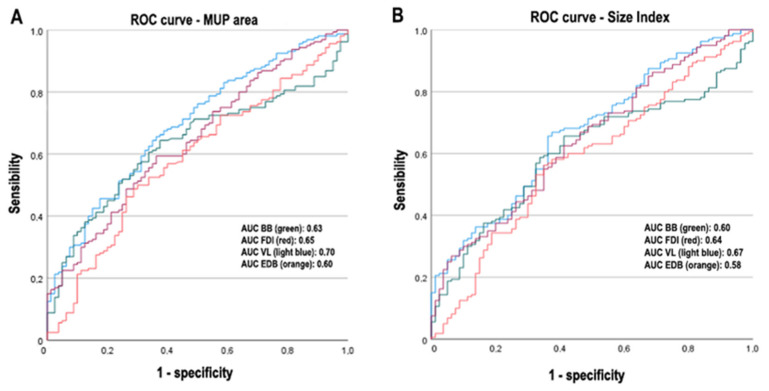
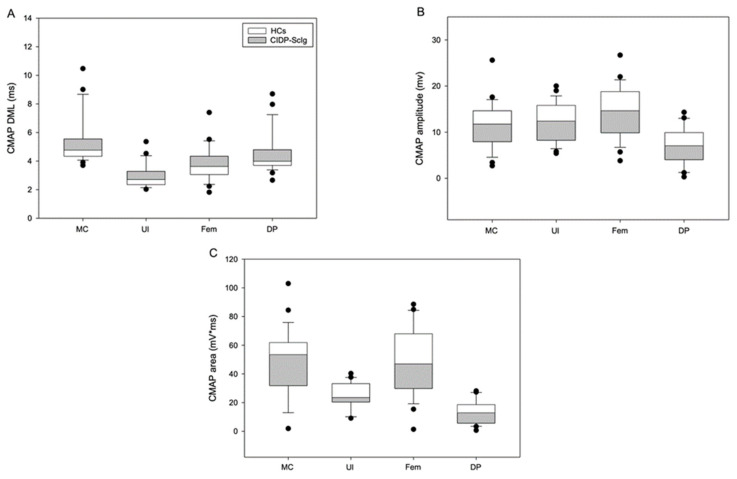
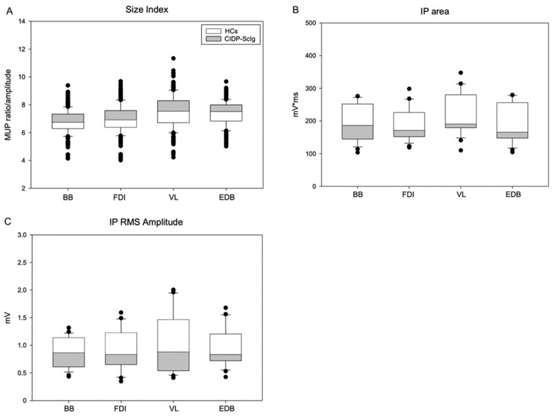
In this work, we aim to identify sensitive neurophysiological biomarkers of axonal degeneration in CIDP patients. A total of 16 CIDP patients, fulfilling the clinical and neurophysiological criteria for typical CIDP, treated with subcutaneous immunoglobulin (ScIg) (0.4 g/kg/week) were evaluated at baseline (before ScIg treatment) and after long-term treatment with ScIg (24 months) by clinical assessment scales, nerve conduction studies (NCS) and electromyography (EMG). Conventional and non-conventional neurophysiological parameters: motor unit potential (MUP) analysis, MUP thickness and size index (SI)] and interference pattern (IP) features were evaluated after long-term treatment (24 months) and compared with a population of 16 healthy controls (HC). An increase of distal motor latency (DML) and reduced compound motor action potential (CMAP) amplitude and area in CIDP patients suggest axonal damage of motor fibers, together with a significant increase of MUP amplitude, duration and area. Analysis of non-conventional MUP parameters shows no difference for MUP thickness; however, in CIDP patients, SI is increased and IP area and amplitude values are lower than HC. Despite clinical and neurophysiological improvement after ScIg treatment, neurophysiological analysis revealed axonal degeneration of motor fibers and motor unit remodeling. Correlation analysis shows that the axonal degeneration process is related to the diagnostic and therapeutic delay. MUP area and SI parameters can detect early signs of axonal degeneration, and their introduction in clinical practice may help to identify patients with the worst outcome.
在这项研究中,我们旨在识别慢性炎性脱髓鞘性多发性神经根神经病(CIDP)患者轴突退变的敏感神经生理学生物标志物。共有16例符合典型CIDP临床和神经生理学标准的患者,接受皮下注射免疫球蛋白(ScIg)(0.4 g/kg/周)治疗,在基线期(ScIg治疗前)以及接受ScIg长期治疗(24个月)后,通过临床评估量表、神经传导研究(NCS)和肌电图(EMG)进行评估。在长期治疗(24个月)后,评估常规和非常规神经生理学参数:运动单位电位(MUP)分析、MUP厚度和大小指数(SI)以及干扰图(IP)特征,并与16名健康对照者(HC)群体进行比较。CIDP患者远端运动潜伏期(DML)增加、复合运动动作电位(CMAP)幅度和面积减小,提示运动纤维存在轴突损伤,同时MUP幅度、时限和面积显著增加。非常规MUP参数分析显示MUP厚度无差异;然而,在CIDP患者中,SI增加,IP面积和幅度值低于HC。尽管ScIg治疗后临床和神经生理学有所改善,但神经生理学分析显示运动纤维存在轴突退变和运动单位重塑。相关性分析表明,轴突退变过程与诊断和治疗延迟有关。MUP面积和SI参数可检测轴突退变的早期迹象,将其引入临床实践可能有助于识别预后最差的患者。