• 文献检索
  • 文档翻译
  • 深度研究
  • 学术资讯
  • Suppr Zotero 插件Zotero 插件
  • 邀请有礼
  • 套餐&价格
  • 历史记录
应用&插件
Suppr Zotero 插件Zotero 插件浏览器插件Mac 客户端Windows 客户端微信小程序
定价
高级版会员购买积分包购买API积分包
服务
文献检索文档翻译深度研究API 文档MCP 服务
关于我们
关于 Suppr公司介绍联系我们用户协议隐私条款
关注我们

Suppr 超能文献

核心技术专利:CN118964589B侵权必究
粤ICP备2023148730 号-1Suppr @ 2026

文献检索

告别复杂PubMed语法,用中文像聊天一样搜索,搜遍4000万医学文献。AI智能推荐,让科研检索更轻松。

立即免费搜索

文件翻译

保留排版,准确专业,支持PDF/Word/PPT等文件格式,支持 12+语言互译。

免费翻译文档

深度研究

AI帮你快速写综述,25分钟生成高质量综述,智能提取关键信息,辅助科研写作。

立即免费体验

GDNF 通过激活 RET 通路增加海马体主神经元的抑制性突触传递。

GDNF Increases Inhibitory Synaptic Drive on Principal Neurons in the Hippocampus via Activation of the Ret Pathway.

机构信息

Experimental Epilepsy Group, Department of Clinical Sciences, Faculty of Medicine, Lund University, Sölvegatan 17, BMC A11, 22362 Lund, Sweden.

Epilepsy Center, Department of Clinical Sciences, Faculty of Medicine, Lund University, Sölvegatan 17, BMC A11, 22362 Lund, Sweden.

出版信息

Int J Mol Sci. 2022 Oct 29;23(21):13190. doi: 10.3390/ijms232113190.

DOI:10.3390/ijms232113190
PMID:36361981
原文链接:https://pmc.ncbi.nlm.nih.gov/articles/PMC9653719/
Abstract

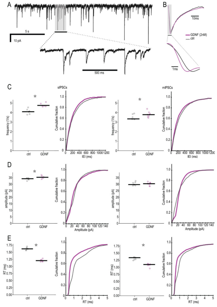
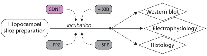
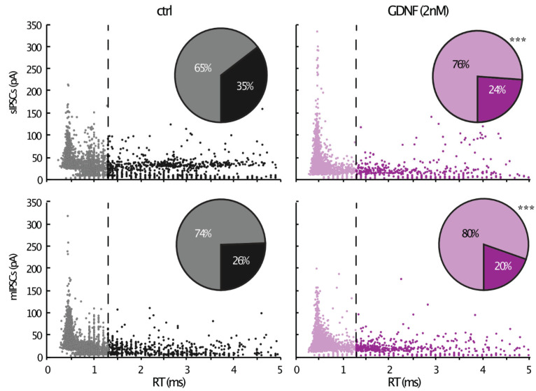
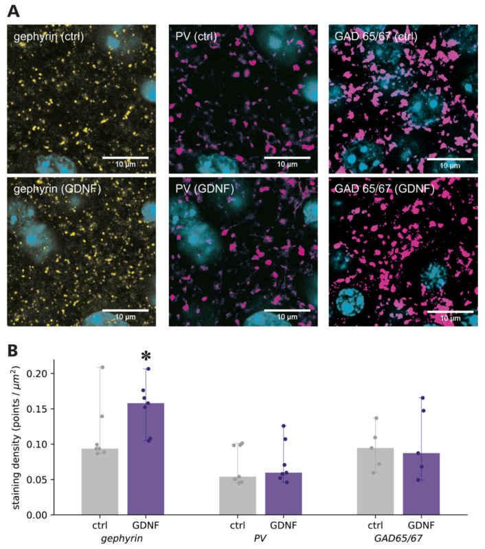
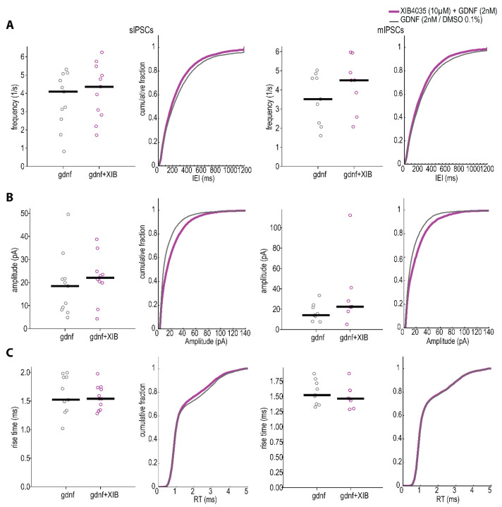
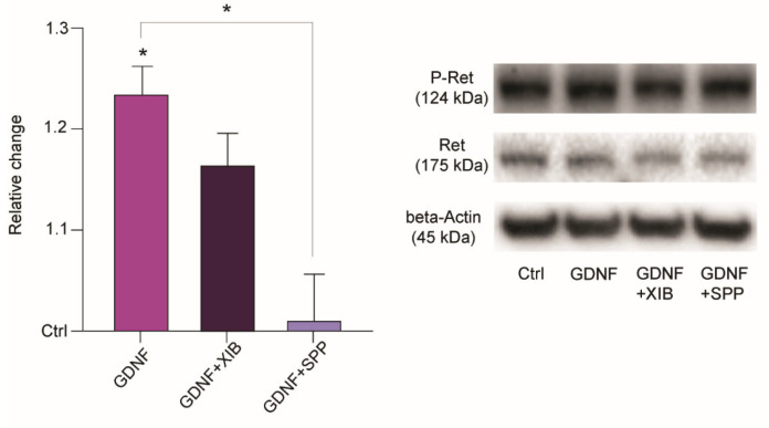
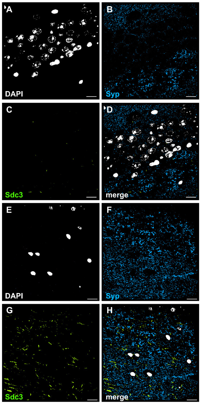
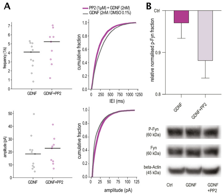
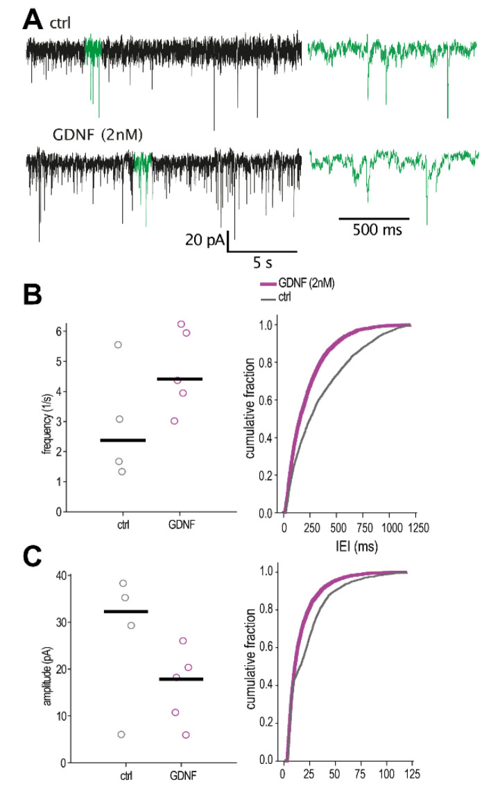
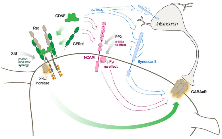
Glial cell line-derived neurotrophic factor (GDNF) has been shown to counteract seizures when overexpressed or delivered into the brain in various animal models of epileptogenesis or chronic epilepsy. The mechanisms underlying this effect have not been investigated. We here demonstrate for the first time that GDNF enhances GABAergic inhibitory drive onto mouse pyramidal neurons by modulating postsynaptic GABA receptors, particularly in perisomatic inhibitory synapses, by GFRα1 mediated activation of the Ret receptor pathway. Other GDNF receptors, such as NCAM or Syndecan3, are not contributing to this effect. We observed similar alterations by GDNF in human hippocampal slices resected from epilepsy patients. These data indicate that GDNF may exert its seizure-suppressant action by enhancing GABAergic inhibitory transmission in the hippocampal network, thus counteracting the increased excitability of the epileptic brain. This new knowledge can contribute to the development of novel, more precise treatment strategies based on a GDNF gene therapy approach.

摘要

胶质细胞源性神经营养因子(GDNF)在各种癫痫发生或慢性癫痫的动物模型中过度表达或递送至大脑时,已被证明可对抗癫痫发作。但其作用机制尚未被研究。我们首次证明,GDNF 通过 GFRα1 介导的 Ret 受体途径的激活,调节突触后 GABA 受体,特别是在体周抑制性突触上,增强 GABA 能抑制性驱动至小鼠锥体神经元。其他 GDNF 受体,如 NCAM 或 Syndecan3,对这种作用没有贡献。我们在从癫痫患者切除的人类海马切片中观察到 GDNF 产生了类似的改变。这些数据表明,GDNF 可能通过增强海马网络中的 GABA 能抑制性传递来发挥其抑制癫痫发作的作用,从而抵消癫痫大脑的过度兴奋。这一新知识可以为基于 GDNF 基因治疗方法的新型、更精确的治疗策略的发展做出贡献。

https://cdn.ncbi.nlm.nih.gov/pmc/blobs/0675/9653719/36999f766c6c/ijms-23-13190-g010.jpg
https://cdn.ncbi.nlm.nih.gov/pmc/blobs/0675/9653719/c4c578090630/ijms-23-13190-g001.jpg
https://cdn.ncbi.nlm.nih.gov/pmc/blobs/0675/9653719/69637745ad0b/ijms-23-13190-g002.jpg
https://cdn.ncbi.nlm.nih.gov/pmc/blobs/0675/9653719/e6ba023eee82/ijms-23-13190-g003.jpg
https://cdn.ncbi.nlm.nih.gov/pmc/blobs/0675/9653719/cd630c501316/ijms-23-13190-g004.jpg
https://cdn.ncbi.nlm.nih.gov/pmc/blobs/0675/9653719/31b62c6e541a/ijms-23-13190-g005.jpg
https://cdn.ncbi.nlm.nih.gov/pmc/blobs/0675/9653719/f807211322ba/ijms-23-13190-g006.jpg
https://cdn.ncbi.nlm.nih.gov/pmc/blobs/0675/9653719/061068cae3cd/ijms-23-13190-g007.jpg
https://cdn.ncbi.nlm.nih.gov/pmc/blobs/0675/9653719/e40de5d661cf/ijms-23-13190-g008.jpg
https://cdn.ncbi.nlm.nih.gov/pmc/blobs/0675/9653719/d7bb6575ec39/ijms-23-13190-g009.jpg
https://cdn.ncbi.nlm.nih.gov/pmc/blobs/0675/9653719/36999f766c6c/ijms-23-13190-g010.jpg

相似文献

1
GDNF Increases Inhibitory Synaptic Drive on Principal Neurons in the Hippocampus via Activation of the Ret Pathway.GDNF 通过激活 RET 通路增加海马体主神经元的抑制性突触传递。
Int J Mol Sci. 2022 Oct 29;23(21):13190. doi: 10.3390/ijms232113190.
2
Down-regulation of microRNA-216a confers protection against yttrium aluminium garnet laser-induced retinal injury the GDNF-mediated GDNF/GFRα1/RET signalling pathway.微小RNA-216a的下调通过胶质细胞源性神经营养因子介导的GDNF/GFRα1/RET信号通路赋予对钇铝石榴石激光诱导的视网膜损伤的保护作用。
J Biosci. 2018 Dec;43(5):985-1000.
3
GFRα1 released by nerves enhances cancer cell perineural invasion through GDNF-RET signaling.神经释放的 GFRα1 通过 GDNF-RET 信号增强癌细胞的神经周围浸润。
Proc Natl Acad Sci U S A. 2014 May 13;111(19):E2008-17. doi: 10.1073/pnas.1402944111. Epub 2014 Apr 28.
4
RET-mediated glial cell line-derived neurotrophic factor signaling inhibits mouse prostate development.RET介导的胶质细胞源性神经营养因子信号传导抑制小鼠前列腺发育。
Development. 2017 Jun 15;144(12):2282-2293. doi: 10.1242/dev.145086. Epub 2017 May 15.
5
Zebrafish GDNF and its co-receptor GFRα1 activate the human RET receptor and promote the survival of dopaminergic neurons in vitro.斑马鱼胶质细胞源性神经营养因子(GDNF)及其共受体GFRα1激活人类RET受体,并在体外促进多巴胺能神经元的存活。
PLoS One. 2017 May 3;12(5):e0176166. doi: 10.1371/journal.pone.0176166. eCollection 2017.
6
SorLA controls neurotrophic activity by sorting of GDNF and its receptors GFRα1 and RET.SorLA 通过对 GDNF 及其受体 GFRα1 和 RET 的分拣来控制神经营养活性。
Cell Rep. 2013 Jan 31;3(1):186-99. doi: 10.1016/j.celrep.2012.12.011. Epub 2013 Jan 17.
7
MET signaling in GABAergic neuronal precursors of the medial ganglionic eminence restricts GDNF activity in cells that express GFRα1 and a new transmembrane receptor partner.中脑内侧神经节隆起的 GABA 能神经元前体细胞中的 MET 信号限制了表达 GFRα1 和新跨膜受体伴侣的细胞中的 GDNF 活性。
J Cell Sci. 2011 Aug 15;124(Pt 16):2797-805. doi: 10.1242/jcs.083717.
8
GM1 ganglioside enhances Ret signaling in striatum.GM1神经节苷脂增强纹状体中的Ret信号传导。
J Neurochem. 2014 Aug;130(4):541-54. doi: 10.1111/jnc.12760. Epub 2014 Jun 13.
9
Expression of GDNF receptors GFRalpha1 and RET is preserved in substantia nigra pars compacta of aging Asian Indians.在衰老的亚洲印第安人中,GDNF 受体 GFRalpha1 和 RET 的表达在黑质致密部中得以保留。
J Chem Neuroanat. 2010 Sep;40(1):43-52. doi: 10.1016/j.jchemneu.2010.03.007. Epub 2010 Mar 27.
10
The GDNF-GFRα1 complex promotes the development of hippocampal dendritic arbors and spines via NCAM.胶质细胞源性神经营养因子-胶质细胞源性神经营养因子受体α1复合物通过神经细胞黏附分子促进海马树突棘和树突分支的发育。
Development. 2016 Nov 15;143(22):4224-4235. doi: 10.1242/dev.140350. Epub 2016 Oct 5.

引用本文的文献

1
Exploring the diversity of biological processes regulated by glial cell line-derived neurotrophic factor, a pleiotropic molecule with therapeutic potential.探索由胶质细胞系衍生的神经营养因子所调控的生物过程的多样性,这是一种具有治疗潜力的多效性分子。
Front Physiol. 2025 Jun 26;16:1618330. doi: 10.3389/fphys.2025.1618330. eCollection 2025.
2
Effects of oxidative stress and GDNF on patients with bipolar disorder: a prospective study.氧化应激和胶质细胞源性神经营养因子对双相情感障碍患者的影响:一项前瞻性研究。
BMC Psychiatry. 2025 Mar 19;25(1):258. doi: 10.1186/s12888-025-06698-3.
3
Neuroprotective and inflammatory biomarkers in pediatric drug-resistant epilepsy: Interplay between GDNF, IL-1β and vitamin D 25-OH.

本文引用的文献

1
Lipid mediator n-3 docosapentaenoic acid-derived protectin D1 enhances synaptic inhibition of hippocampal principal neurons by interaction with a G-protein-coupled receptor.脂质介质 n-3 二十二碳五烯酸衍生的保护素 D1 通过与 G 蛋白偶联受体相互作用增强海马主神经元的突触抑制。
FASEB J. 2022 Mar;36(3):e22203. doi: 10.1096/fj.202101815R.
2
Comparison of biophysical properties of α1β2 and α3β2 GABAA receptors in whole-cell patch-clamp electrophysiological recordings.全细胞膜片钳电生理记录中 α1β2 和 α3β2 GABAA 受体的生物物理特性比较。
PLoS One. 2020 Jun 1;15(6):e0234080. doi: 10.1371/journal.pone.0234080. eCollection 2020.
3
小儿耐药性癫痫中的神经保护和炎症生物标志物:胶质细胞源性神经营养因子、白细胞介素-1β和25-羟基维生素D之间的相互作用
Narra J. 2024 Dec;4(3):e1581. doi: 10.52225/narra.v4i3.1581. Epub 2024 Nov 18.
4
Reduced Levels of Lacrimal Glial Cell Line-Derived Neurotrophic Factor (GDNF) in Patients with Focal Epilepsy and Focal Epilepsy with Comorbid Depression: A Biomarker Candidate.泪腺神经胶质细胞衍生神经营养因子(GDNF)水平降低与局灶性癫痫和伴发抑郁的局灶性癫痫患者相关:一个生物标志物候选者。
Int J Mol Sci. 2023 Nov 27;24(23):16818. doi: 10.3390/ijms242316818.
Inhibition of epileptiform activity by neuropeptide Y in brain tissue from drug-resistant temporal lobe epilepsy patients.
神经肽 Y 抑制耐药性颞叶癫痫患者脑组织中的癫痫样活动。
Sci Rep. 2019 Dec 18;9(1):19393. doi: 10.1038/s41598-019-56062-1.
4
Long-Term, Targeted Delivery of GDNF from Encapsulated Cells Is Neuroprotective and Reduces Seizures in the Pilocarpine Model of Epilepsy.包封细胞长效、靶向递送 GDNF 可保护神经并减少匹罗卡品癫痫模型中的癫痫发作。
J Neurosci. 2019 Mar 13;39(11):2144-2156. doi: 10.1523/JNEUROSCI.0435-18.2018. Epub 2019 Jan 21.
5
Unilateral ex vivo gene therapy by GDNF in epileptic rats.癫痫大鼠单侧离体基因治疗 GDNF。
Gene Ther. 2019 Apr;26(3-4):65-74. doi: 10.1038/s41434-018-0050-7. Epub 2018 Nov 21.
6
Prolonged life of human acute hippocampal slices from temporal lobe epilepsy surgery.从颞叶癫痫手术中获取的人类急性海马脑片的延长寿命。
Sci Rep. 2018 Mar 7;8(1):4158. doi: 10.1038/s41598-018-22554-9.
7
Treatment Outcomes in Patients With Newly Diagnosed Epilepsy Treated With Established and New Antiepileptic Drugs: A 30-Year Longitudinal Cohort Study.新诊断癫痫患者使用现有和新型抗癫痫药物治疗的治疗结局:一项长达 30 年的纵向队列研究。
JAMA Neurol. 2018 Mar 1;75(3):279-286. doi: 10.1001/jamaneurol.2017.3949.
8
Compromised Survival of Cerebellar Molecular Layer Interneurons Lacking GDNF Receptors GFRα1 or RET Impairs Normal Cerebellar Motor Learning.缺乏胶质细胞源性神经营养因子受体GFRα1或RET的小脑分子层中间神经元存活受损,会损害正常的小脑运动学习。
Cell Rep. 2017 Jun 6;19(10):1977-1986. doi: 10.1016/j.celrep.2017.05.030.
9
Bayesian Inference of Synaptic Quantal Parameters from Correlated Vesicle Release.基于相关囊泡释放的突触量子参数的贝叶斯推断
Front Comput Neurosci. 2016 Nov 25;10:116. doi: 10.3389/fncom.2016.00116. eCollection 2016.
10
Post-mortem brain analyses of the Lothian Birth Cohort 1936: extending lifetime cognitive and brain phenotyping to the level of the synapse.对洛锡安出生队列 1936 年的死后大脑分析:将终生认知和大脑表型分析扩展到突触水平。
Acta Neuropathol Commun. 2015 Sep 4;3:53. doi: 10.1186/s40478-015-0232-0.