Clinical Pathology Department, Faculty of Veterinary Medicine, Kafrelsheikh University, Kafrelsheikh, 33516, Egypt.
Department of Fish Processing and Biotechnology, Faculty of Aquatic and Fisheries Sciences, Kafrelsheikh University, Kafrelsheikh, 33516, Egypt.
Environ Sci Pollut Res Int. 2023 Feb;30(10):26308-26326. doi: 10.1007/s11356-022-23876-y. Epub 2022 Nov 11.
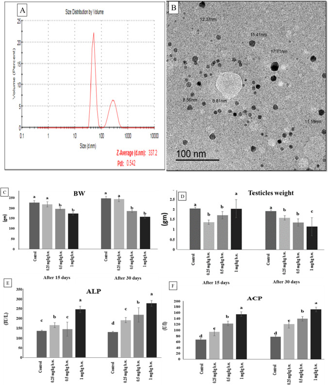
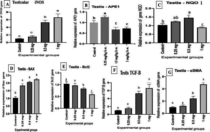
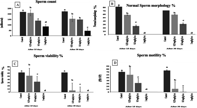
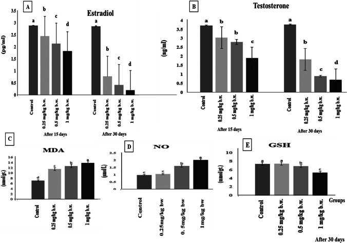
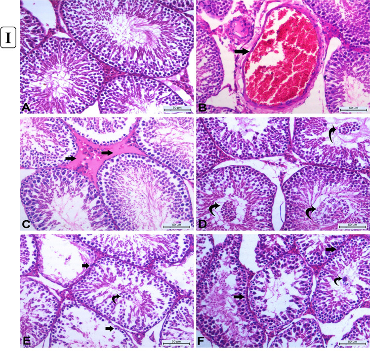
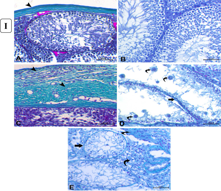
In medicine, silver nanoparticles (AgNPs) are employed often. They do, however, have negative impacts, particularly on the reproductive organs. This research aimed to assess AgNP impact on the testis and the possible intracellular mechanisms to induce testicular deteriorations in rats at various concentrations and different time intervals. Sprague Dawley rats (n = 40) were allocated into four equal groups: the control one, and three other groups injected intra-peritoneally with AgNP solution 0.25, 0.5, and 1 mg/kg b.w. respectively for 15 and 30 days. Our findings revealed that AgNPs reduced body and testicular weights, estradiol (E2) and testosterone (T) hormone levels, and sperm parameters while elevating the nitric oxide and malondialdehyde levels with inhibition of reduced glutathione contents in testicular tissue. Interestingly, AgNPs significantly upregulated the testicular inducible nitric oxide synthase, B cell lymphoma 2 (Bcl-2)-associated X, transforming growth factor, and alpha-smooth muscle actin (α-SMA) expression levels. However, apurinic/apyrimidinic endo deoxyribonuclease 1 (APE1), NAD (P) H quinone dehydrogenase 1 (NQO1), and Bcl-2 expression levels were all downregulated indicating exhaustion of body antioxidant and repairing defense mechanisms in testicles in comparison with the control rats. Various histological alterations were also detected which dramatically increased in rats sacrificed after 30 days such as loss of the lining cells of seminiferous tubules with no spermatozoa and tubular irregularities associated with thickening of their basement membranes. Immunolabeling implicated in the apoptotic pathway revealed a negative expression of Bcl-2 and marked immunoreactivity for caspase-3 after 30 days of AgNP treatment in comparison to the control rats. To our knowledge, there have been no previous publications on the role of the α-SMA, APE1, and NQO1 genes in the molecular pathogenesis of AgNP testicular cytotoxicity following AgNP acute and chronic exposure.
在医学领域,经常使用银纳米粒子(AgNPs)。然而,它们确实有负面影响,特别是对生殖器官。本研究旨在评估 AgNP 对不同浓度和不同时间间隔的大鼠睾丸的影响,以及可能导致睾丸损伤的细胞内机制。将 40 只 Sprague Dawley 大鼠分为四组:对照组和另外三组,分别腹腔注射 0.25、0.5 和 1 mg/kg bw 的 AgNP 溶液,持续 15 和 30 天。我们的研究结果表明,AgNPs 降低了体重和睾丸重量、雌二醇(E2)和睾酮(T)激素水平以及精子参数,同时提高了睾丸组织中一氧化氮和丙二醛水平,降低了还原型谷胱甘肽含量。有趣的是,AgNPs 显著上调了睾丸诱导型一氧化氮合酶、B 细胞淋巴瘤 2(Bcl-2)相关 X、转化生长因子和α-平滑肌肌动蛋白(α-SMA)的表达水平。然而,APEN1、NAD(P)H 醌氧化还原酶 1(NQO1)和 Bcl-2 的表达水平均下调,表明与对照组大鼠相比,睾丸中的抗氧化和修复防御机制已经耗尽。还检测到各种组织学改变,在第 30 天处死的大鼠中,这些改变更为明显,如精小管衬里细胞丢失,没有精子,小管不规则,基底膜增厚。免疫标记法表明,在 AgNP 处理 30 天后,与对照组大鼠相比,凋亡途径中的 Bcl-2 表达呈阴性,caspase-3 免疫反应性显著增强。据我们所知,以前没有关于α-SMA、APEN1 和 NQO1 基因在 AgNP 急性和慢性暴露后睾丸细胞毒性的分子发病机制中的作用的出版物。