• 文献检索
  • 文档翻译
  • 深度研究
  • 学术资讯
  • Suppr Zotero 插件Zotero 插件
  • 邀请有礼
  • 套餐&价格
  • 历史记录
应用&插件
Suppr Zotero 插件Zotero 插件浏览器插件Mac 客户端Windows 客户端微信小程序
定价
高级版会员购买积分包购买API积分包
服务
文献检索文档翻译深度研究API 文档MCP 服务
关于我们
关于 Suppr公司介绍联系我们用户协议隐私条款
关注我们

Suppr 超能文献

核心技术专利:CN118964589B侵权必究
粤ICP备2023148730 号-1Suppr @ 2026

文献检索

告别复杂PubMed语法,用中文像聊天一样搜索,搜遍4000万医学文献。AI智能推荐,让科研检索更轻松。

立即免费搜索

文件翻译

保留排版,准确专业,支持PDF/Word/PPT等文件格式,支持 12+语言互译。

免费翻译文档

深度研究

AI帮你快速写综述,25分钟生成高质量综述,智能提取关键信息,辅助科研写作。

立即免费体验

用于扫描电子显微镜(SEM)分析的非导电涂层微样品的导电横截面制备。

Conductive cross-section preparation of non-conductive painting micro-samples for SEM analysis.

作者信息

Jaques Victory Armida Janine, Zikmundová Eva, Holas Jiří, Zikmund Tomáš, Kaiser Jozef, Holcová Katarína

机构信息

Institute of Geology and Palaeontology, Faculty of Science, Charles University, Albertov 6, 12843, Praha 2, Czech Republic.

CEITEC - Central European Institute of Technology, Brno University of Technology, Purkyňova 656/123, 612 00, Brno, Czech Republic.

出版信息

Sci Rep. 2022 Nov 16;12(1):19650. doi: 10.1038/s41598-022-21882-1.

DOI:10.1038/s41598-022-21882-1
PMID:36385138
原文链接:https://pmc.ncbi.nlm.nih.gov/articles/PMC9668980/
Abstract

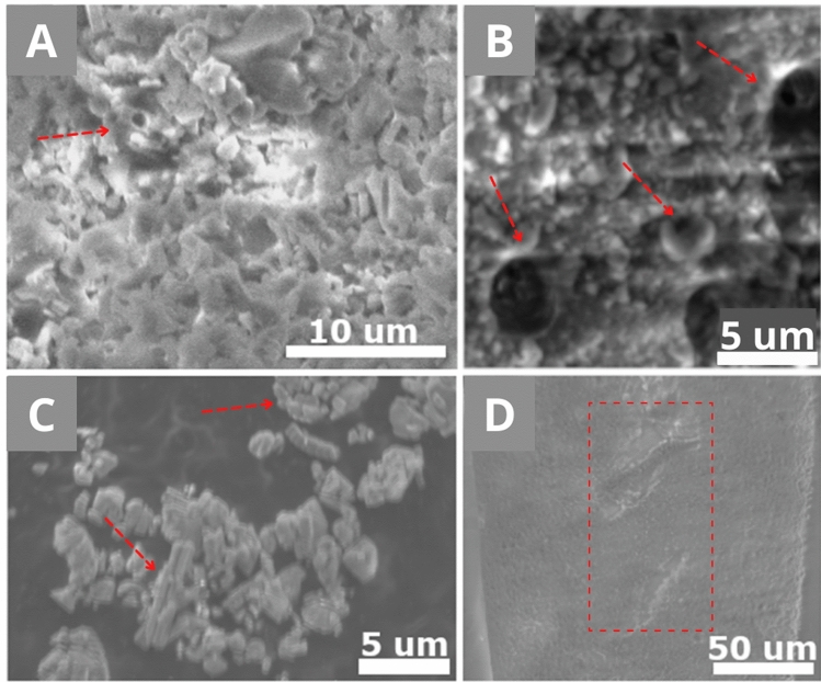
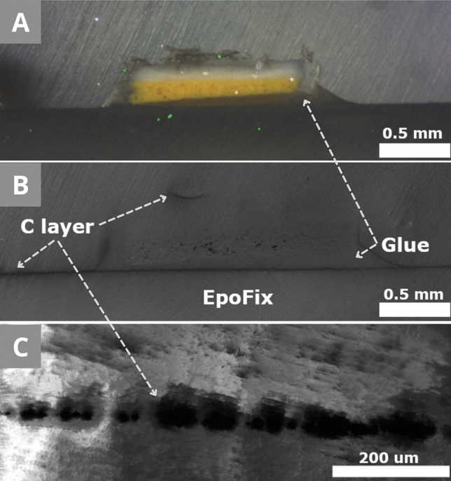
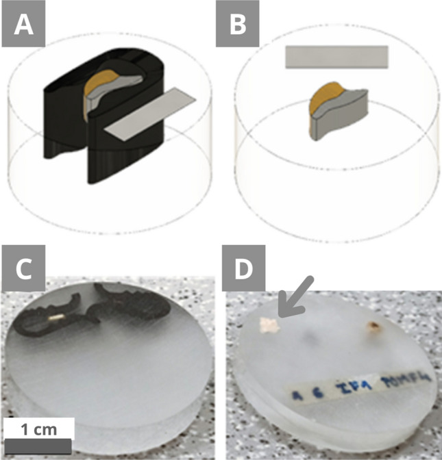
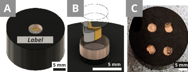
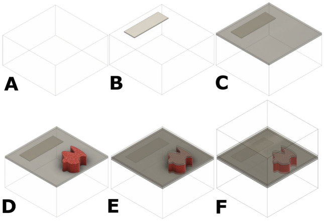
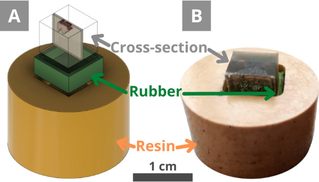
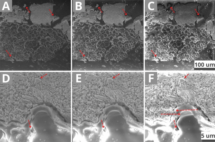
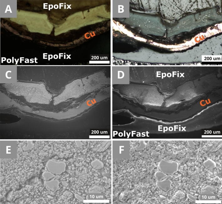
Scanning electron microscopy (SEM) is a common method for the analysis of painting micro-samples. The high resolution of this technique offers precise surface analysis and can be coupled with an energy-dispersive spectrometer for the acquisition of the elemental composition. For light microscopy and SEM analysis, the painting micro-samples are commonly prepared as cross-sections, where the micro-sample positioned on the side is embedded in a resin. Therefore, the sequence of its layers is exposed after the cross-section is polished. In common cases outside of cultural heritage, a conductive layer is applied on the polished side, but in this field, the measurements are mostly done in low-vacuum SEM (LV-SEM). Although the charging effect is reduced in LV-SEM, it can still occur, and can hardly be prevented even with carbon tape or paint. This work presents two conductive cross-section preparation methods for non-conductive samples, which reduce charging effects without impairing the sample integrity.

摘要

扫描电子显微镜(SEM)是分析绘画微样本的常用方法。该技术的高分辨率可提供精确的表面分析,并且可以与能量色散光谱仪联用,以获取元素组成。对于光学显微镜和扫描电子显微镜分析,绘画微样本通常制备成横截面,其中位于侧面的微样本嵌入树脂中。因此,在横截面抛光后,其层序就会暴露出来。在文化遗产领域之外的常见情况下,会在抛光面上施加导电层,但在该领域,测量大多在低真空扫描电子显微镜(LV-SEM)中进行。尽管在低真空扫描电子显微镜中充电效应会降低,但它仍然可能发生,即使使用碳带或涂料也很难防止。这项工作提出了两种用于非导电样本的导电横截面制备方法,它们在不损害样本完整性的情况下减少了充电效应。

https://cdn.ncbi.nlm.nih.gov/pmc/blobs/dfec/9668980/9080431343cc/41598_2022_21882_Fig10_HTML.jpg
https://cdn.ncbi.nlm.nih.gov/pmc/blobs/dfec/9668980/00c97b4e6287/41598_2022_21882_Fig1_HTML.jpg
https://cdn.ncbi.nlm.nih.gov/pmc/blobs/dfec/9668980/32fc42f8c929/41598_2022_21882_Fig2_HTML.jpg
https://cdn.ncbi.nlm.nih.gov/pmc/blobs/dfec/9668980/f2aac4f6ab36/41598_2022_21882_Fig3_HTML.jpg
https://cdn.ncbi.nlm.nih.gov/pmc/blobs/dfec/9668980/069be5e36c75/41598_2022_21882_Fig4_HTML.jpg
https://cdn.ncbi.nlm.nih.gov/pmc/blobs/dfec/9668980/406ab4bcd85a/41598_2022_21882_Fig5_HTML.jpg
https://cdn.ncbi.nlm.nih.gov/pmc/blobs/dfec/9668980/c74717ee4791/41598_2022_21882_Fig6_HTML.jpg
https://cdn.ncbi.nlm.nih.gov/pmc/blobs/dfec/9668980/8fdbbab0833e/41598_2022_21882_Fig7_HTML.jpg
https://cdn.ncbi.nlm.nih.gov/pmc/blobs/dfec/9668980/a50bfc033c77/41598_2022_21882_Fig8_HTML.jpg
https://cdn.ncbi.nlm.nih.gov/pmc/blobs/dfec/9668980/67daa0b71cb4/41598_2022_21882_Fig9_HTML.jpg
https://cdn.ncbi.nlm.nih.gov/pmc/blobs/dfec/9668980/9080431343cc/41598_2022_21882_Fig10_HTML.jpg

相似文献

1
Conductive cross-section preparation of non-conductive painting micro-samples for SEM analysis.用于扫描电子显微镜(SEM)分析的非导电涂层微样品的导电横截面制备。
Sci Rep. 2022 Nov 16;12(1):19650. doi: 10.1038/s41598-022-21882-1.
2
Cyclododecane shaping, sublimation rate and residue analysis for the extraction of painting micro-samples from resin cross-sections.用于从树脂横截面中提取绘画微样本的环十二烷成型、升华速率及残留分析
Sci Rep. 2022 Nov 16;12(1):19654. doi: 10.1038/s41598-022-22448-x.
3
NMR and other molecular and elemental spectroscopies for the characterization of samples from an outdoor mural painting by Siqueiros.用于表征西凯罗斯一幅户外壁画样本的核磁共振及其他分子和元素光谱学方法。
Spectrochim Acta A Mol Biomol Spectrosc. 2022 Jun 5;274:121073. doi: 10.1016/j.saa.2022.121073. Epub 2022 Feb 24.
4
Publisher Correction: Conductive cross-section preparation of non-conductive painting micro-samples for SEM analysis.出版商更正:用于扫描电子显微镜分析的非导电涂料微样品的导电横截面制备。
Sci Rep. 2022 Dec 28;12(1):22487. doi: 10.1038/s41598-022-26752-4.
5
High resolution SEM imaging of gold nanoparticles in cells and tissues.细胞和组织中金纳米颗粒的高分辨率扫描电子显微镜成像。
J Microsc. 2014 Dec;256(3):237-47. doi: 10.1111/jmi.12179. Epub 2014 Sep 16.
6
Enhancement of protein detection on cultural heritage samples after SYPRO™ Ruby staining by optical microscopy and micro-FTIR spectroscopy.通过光学显微镜和微傅里叶变换红外光谱法增强 SYPRO™ Ruby 染色后文化遗产样品上蛋白质的检测。
Spectrochim Acta A Mol Biomol Spectrosc. 2023 Dec 5;302:123067. doi: 10.1016/j.saa.2023.123067. Epub 2023 Jun 22.
7
Conductive resins improve charging and resolution of acquired images in electron microscopic volume imaging.导电树脂可提高电子显微镜体积成像中获取图像的充电和分辨率。
Sci Rep. 2016 Mar 29;6:23721. doi: 10.1038/srep23721.
8
Scanning Electron Microscopy of Bone.骨的扫描电子显微镜检查
Methods Mol Biol. 2019;1914:571-616. doi: 10.1007/978-1-4939-8997-3_31.
9
Minimal resin embedding of SBF-SEM samples reduces charging and facilitates finding a surface-linked region of interest.SBF-SEM样本的最小树脂包埋可减少电荷积累,并有助于找到感兴趣的表面连接区域。
Front Zool. 2023 Aug 29;20(1):29. doi: 10.1186/s12983-023-00507-x.
10
Multispectral and hyperspectral image analysis of elemental and micro-Raman maps of cross-sections from a 16th century painting.对一幅16世纪画作横截面的元素和显微拉曼光谱图进行多光谱和高光谱图像分析。
Anal Chim Acta. 2008 Mar 3;610(1):15-24. doi: 10.1016/j.aca.2007.12.043. Epub 2008 Jan 9.

引用本文的文献

1
Transport of Nanoparticles into Plants and Their Detection Methods.纳米颗粒向植物中的转运及其检测方法。
Nanomaterials (Basel). 2024 Jan 5;14(2):131. doi: 10.3390/nano14020131.
2
Bulk synthesis of conductive non-metallic carbon nanospheres and a 3D printed carrier device for scanning electron microscope calibration.用于扫描电子显微镜校准的导电非金属碳纳米球的批量合成及三维打印载体装置
RSC Adv. 2023 Jul 19;13(32):21882-21889. doi: 10.1039/d3ra03956k.