Instituto Gulbenkian de Ciência, Fundação Calouste Gulbenkian, Rua da Quinta Grande, Portugal.
J Cell Biol. 2023 Feb 6;222(2). doi: 10.1083/jcb.202209052. Epub 2022 Nov 21.
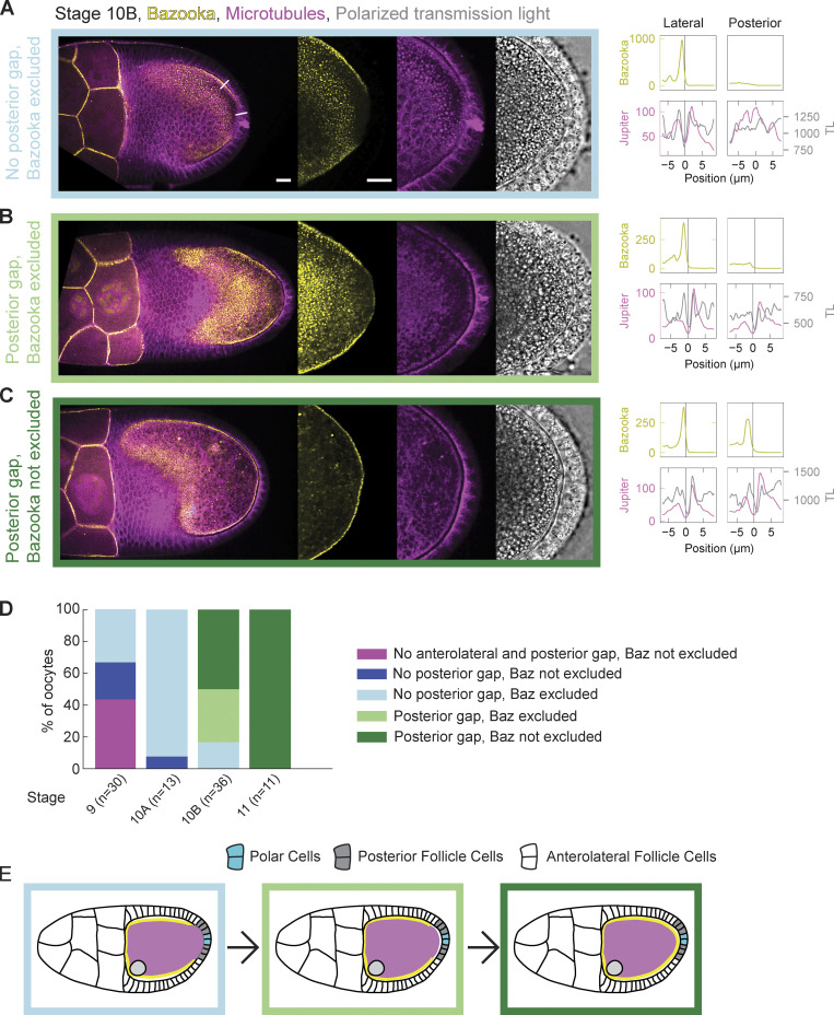
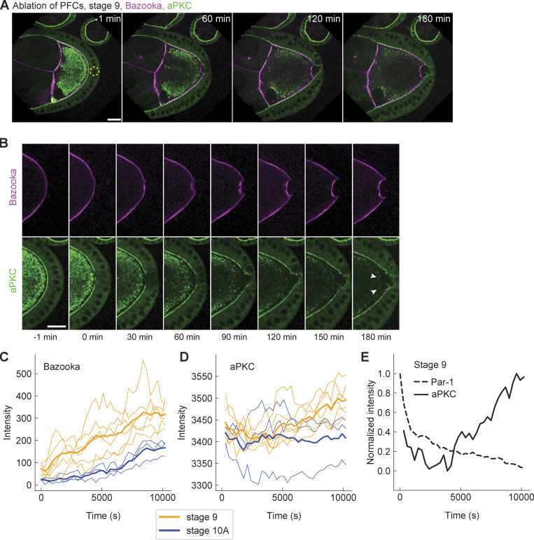
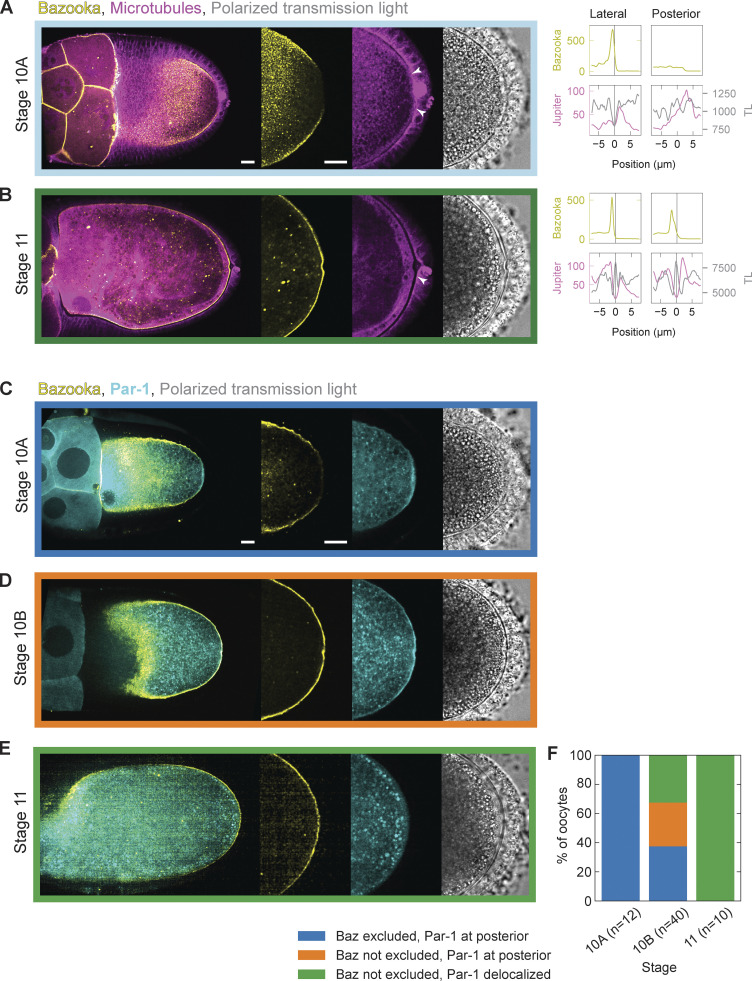
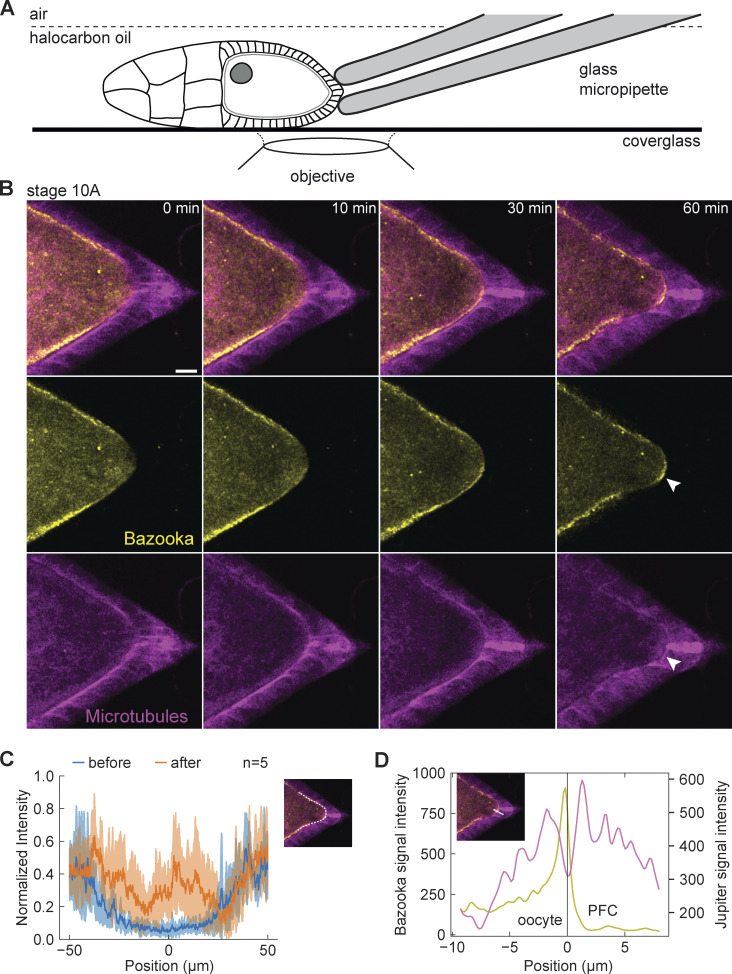
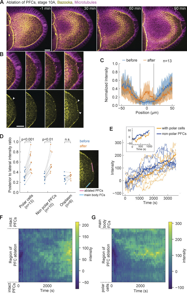
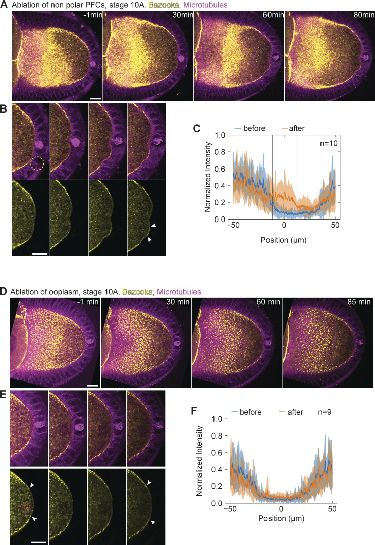
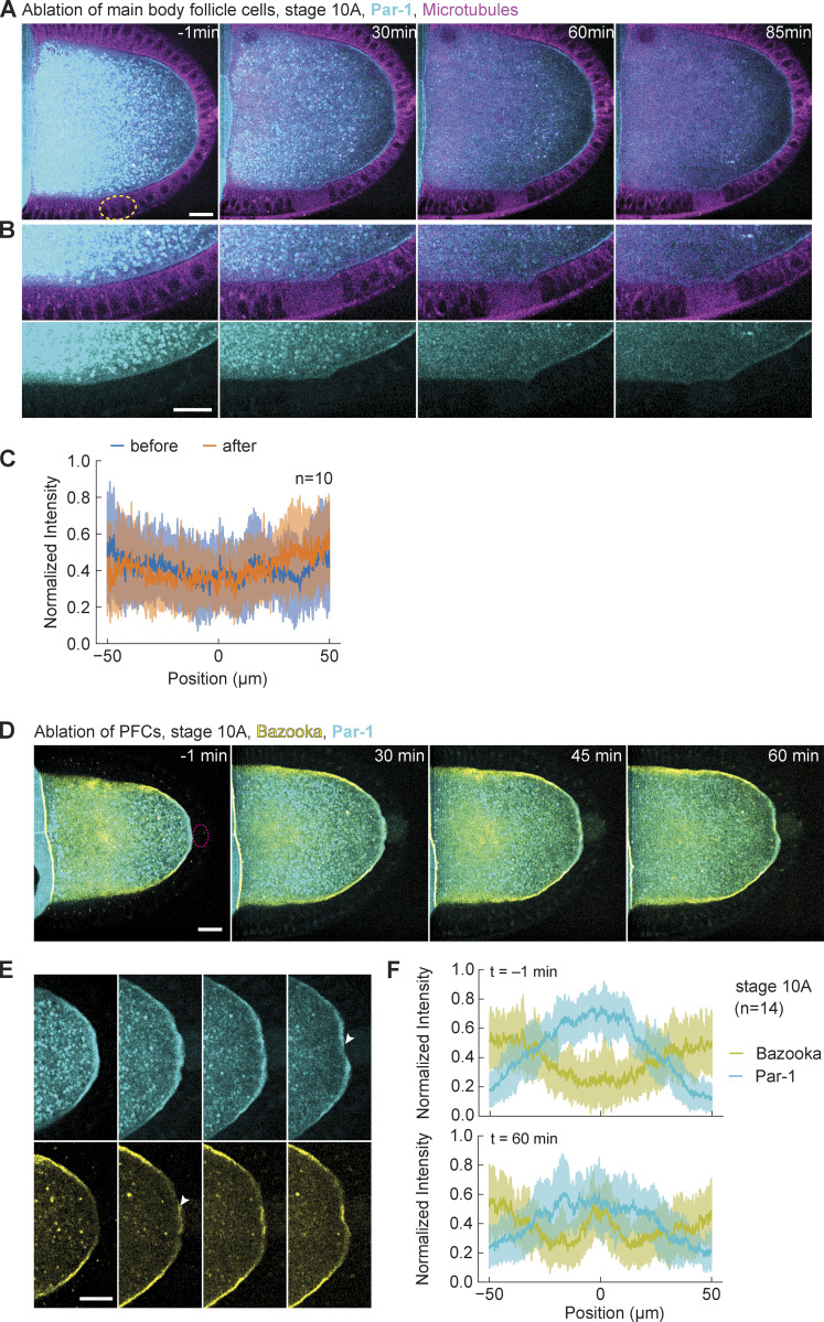
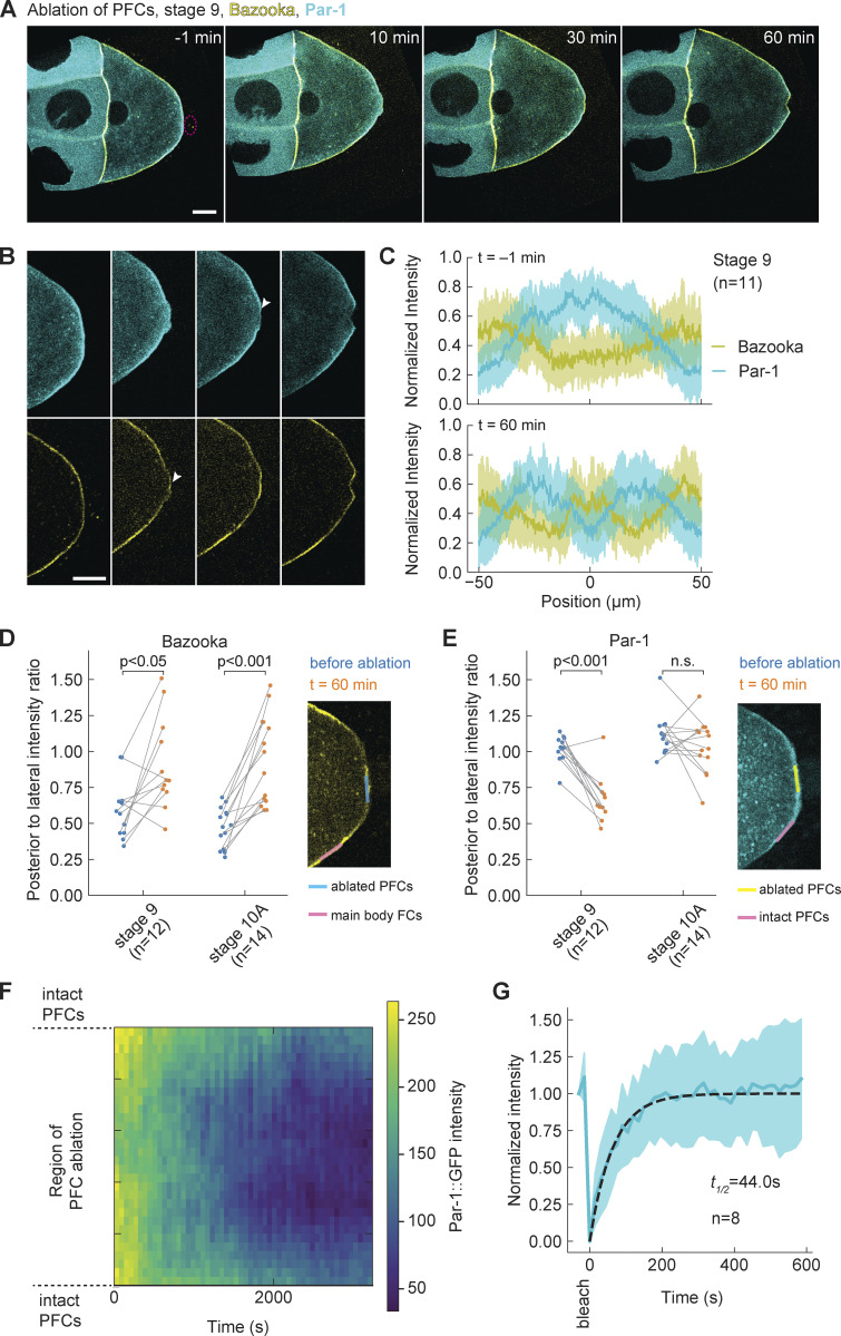
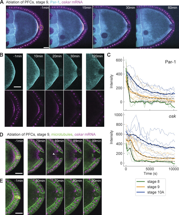
In Drosophila melanogaster, the anterior-posterior body axis is maternally established and governed by differential localization of partitioning defective (Par) proteins within the oocyte. At mid-oogenesis, Par-1 accumulates at the oocyte posterior end, while Par-3/Bazooka is excluded there but maintains its localization along the remaining oocyte cortex. Past studies have proposed the need for somatic cells at the posterior end to initiate oocyte polarization by providing a trigger signal. To date, neither the molecular identity nor the nature of the signal is known. Here, we provide evidence that mechanical contact of posterior follicle cells (PFCs) with the oocyte cortex causes the posterior exclusion of Bazooka and maintains oocyte polarity. We show that Bazooka prematurely accumulates exclusively where posterior follicle cells have been mechanically detached or ablated. Furthermore, we provide evidence that PFC contact maintains Par-1 and oskar mRNA localization and microtubule cytoskeleton polarity in the oocyte. Our observations suggest that cell-cell contact mechanics modulates Par protein binding sites at the oocyte cortex.
在黑腹果蝇中,前后体轴由母体建立,并受卵母细胞内分隔缺陷(Par)蛋白的差异定位控制。在卵母细胞发生的中期,Par-1 积累在卵母细胞的后端,而 Par-3/Bazooka 则被排斥在那里,但仍沿剩余的卵母细胞皮层定位。过去的研究提出需要后端的体细胞通过提供触发信号来启动卵母细胞极化。迄今为止,尚不清楚信号的分子身份或性质。在这里,我们提供的证据表明,后滤泡细胞(PFCs)与卵母细胞皮层的机械接触导致 Bazooka 被排斥在后端,并维持卵母细胞的极性。我们发现,Bazooka 仅在前滤泡细胞被机械分离或消融的地方过早积累。此外,我们提供的证据表明,PFC 接触维持了 Par-1 和 Oskar mRNA 在卵母细胞中的定位和微管细胞骨架极性。我们的观察表明,细胞-细胞接触力学调节了卵母细胞皮层上 Par 蛋白结合位点。