Hier Daniel B, Azizi Sima, Thimgan Matthew S, Wunsch Donald C
Applied Computational Intelligence Laboratory, Department of Electrical & Computer Engineering, Missouri University of Science & Technology, Rolla, MO, United States.
Department of Neurology and Rehabilitation, University of Illinois at Chicago, Chicago, IL, United States.
Front Aging Neurosci. 2022 Nov 9;14:1055170. doi: 10.3389/fnagi.2022.1055170. eCollection 2022.
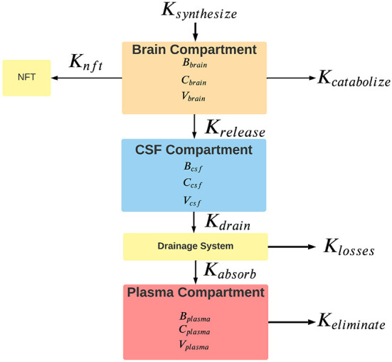
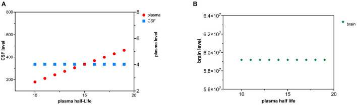
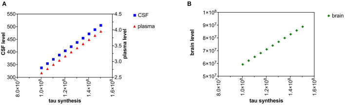
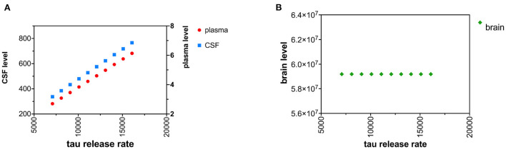
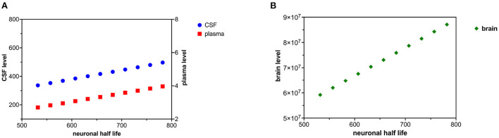
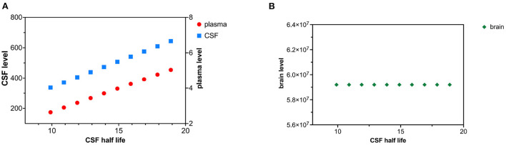
The cytoskeletal protein tau is implicated in the pathogenesis of Alzheimer's disease which is characterized by intra-neuronal neurofibrillary tangles containing abnormally phosphorylated insoluble tau. Levels of soluble tau are elevated in the brain, the CSF, and the plasma of patients with Alzheimer's disease. To better understand the causes of these elevated levels of tau, we propose a three-compartment kinetic model (brain, CSF, and plasma). The model assumes that the synthesis of tau follows zero-order kinetics (uncorrelated with compartmental tau levels) and that the release, absorption, and clearance of tau is governed by first-order kinetics (linearly related to compartmental tau levels). Tau that is synthesized in the brain compartment can be released into the interstitial fluid, catabolized, or retained in neurofibrillary tangles. Tau released into the interstitial fluid can mix with the CSF and eventually drain to the plasma compartment. However, losses of tau in the drainage pathways may be significant. The kinetic model estimates half-life of tau in each compartment (552 h in the brain, 9.9 h in the CSF, and 10 h in the plasma). The kinetic model predicts that an increase in the neuronal tau synthesis rate or a decrease in tau catabolism rate best accounts for observed increases in tau levels in the brain, CSF, and plasma found in Alzheimer's disease. Furthermore, the model predicts that increases in brain half-life of tau in Alzheimer's disease should be attributed to decreased tau catabolism and not to increased tau synthesis. Most clearance of tau in the neuron occurs through catabolism rather than release to the CSF compartment. Additional experimental data would make ascertainment of the model parameters more precise.
细胞骨架蛋白tau与阿尔茨海默病的发病机制有关,该病的特征是神经元内出现含有异常磷酸化不溶性tau的神经原纤维缠结。在阿尔茨海默病患者的大脑、脑脊液和血浆中,可溶性tau水平升高。为了更好地理解tau水平升高的原因,我们提出了一个三室动力学模型(大脑、脑脊液和血浆)。该模型假设tau的合成遵循零级动力学(与各室tau水平无关),而tau的释放、吸收和清除受一级动力学控制(与各室tau水平呈线性相关)。在脑室中合成的tau可以释放到细胞间液中,被分解代谢,或保留在神经原纤维缠结中。释放到细胞间液中的tau可以与脑脊液混合,并最终引流到血浆室。然而,在引流途径中tau的损失可能很大。动力学模型估计了每个室中tau的半衰期(大脑中为552小时,脑脊液中为9.9小时,血浆中为10小时)。该动力学模型预测,神经元tau合成速率的增加或tau分解代谢速率的降低最能解释在阿尔茨海默病患者大脑、脑脊液和血浆中观察到的tau水平升高。此外,该模型预测,阿尔茨海默病中tau在大脑中的半衰期增加应归因于tau分解代谢的减少,而不是tau合成的增加。神经元中tau的大部分清除是通过分解代谢而不是释放到脑脊液室中进行的。更多的实验数据将使模型参数的确定更加精确。