DeWitt Daughtry Family Department of Surgery, University of Miami Leonard M. Miller School of Medicine, Miami, United States.
Department of Medicine, Division of Pulmonary, Critical Care and Sleep, University of Miami, Miami, United States.
Elife. 2022 Dec 1;11:e79543. doi: 10.7554/eLife.79543.
MicroRNAs (miRNA) and other components contained in extracellular vesicles may reflect the presence of a disease. Lung tissue, sputum, and sera of individuals with idiopathic pulmonary fibrosis (IPF) show alterations in miRNA expression. We designed this study to test whether urine and/or tissue derived exosomal miRNAs from individuals with IPF carry cargo that can promote fibrosis.
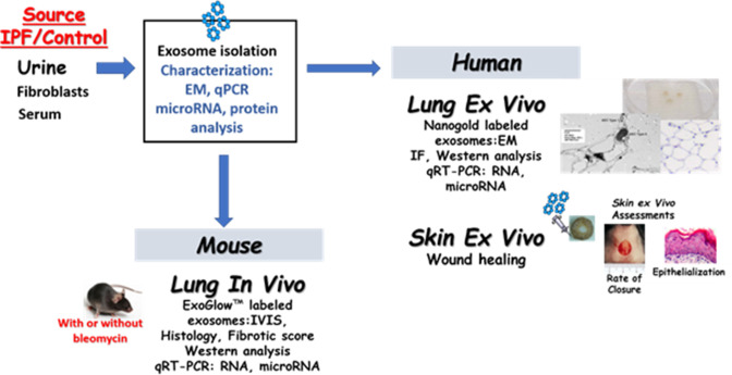
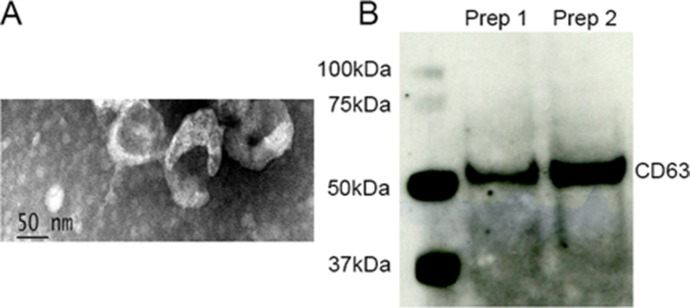
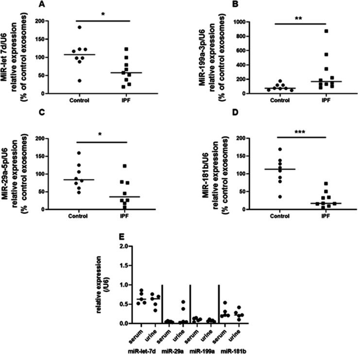
Exosomes were isolated from urine (U-IPFexo), lung tissue myofibroblasts (MF-IPFexo), serum from individuals with IPF (n=16) and age/sex-matched controls without lung disease (n=10). We analyzed microRNA expression of isolated exosomes and their in vivo bio-distribution. We investigated the effect on ex vivo skin wound healing and in in vivo mouse lung models.
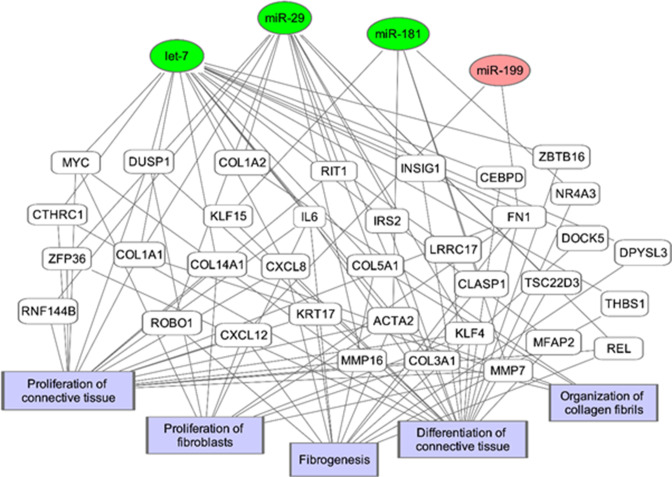
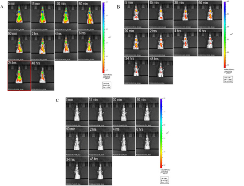
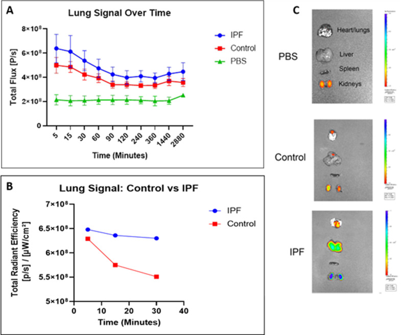
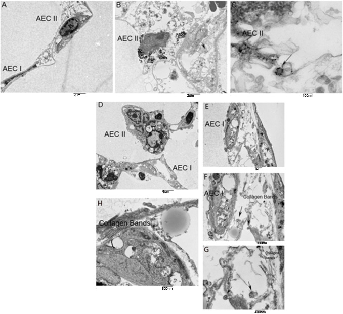
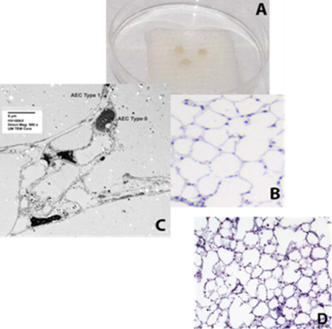
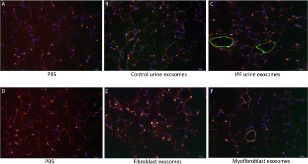
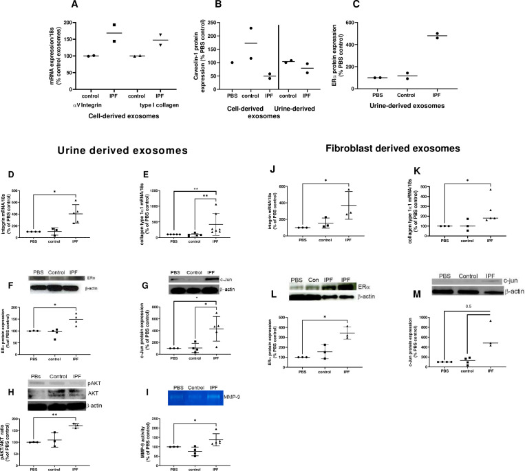
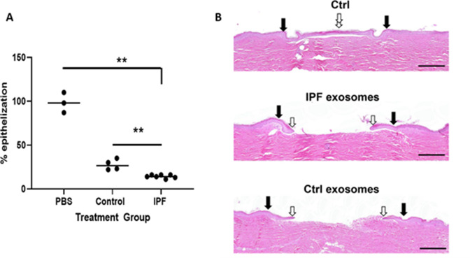
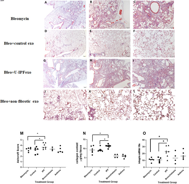
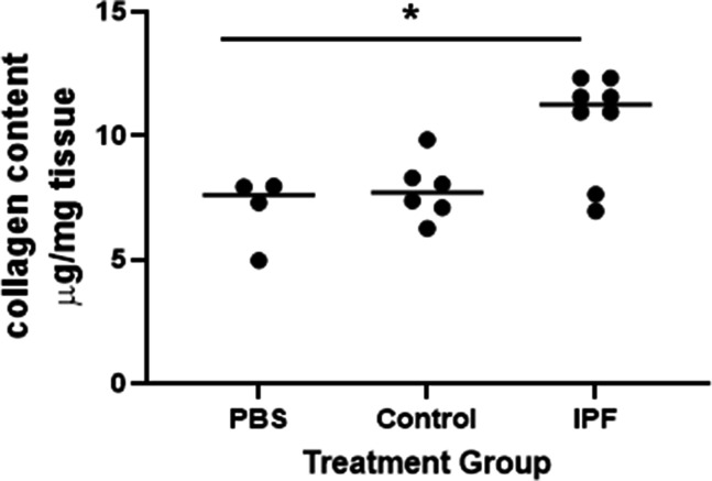
U-IPFexo or MF-IPFexo expressed consistent with previous reports of miRNA expression obtained from lung tissue/sera from patients with IPF. In vivo bio-distribution experiments detected bioluminescent exosomes in the lung of normal C57Bl6 mice within 5 min after intravenous infusion, followed by distribution to other organs irrespective of exosome source. Exosomes labeled with gold nanoparticles and imaged by transmission electron microscopy were visualized in alveolar epithelial type I and type II cells. Treatment of human and mouse lung punches obtained from control, non-fibrotic lungs with either U-IPFexo or MF-IPFexo produced a fibrotic phenotype. A fibrotic phenotype was also induced in a human ex vivo skin model and in in vivo lung models.
Our results provide evidence of a systemic feature of IPF whereby exosomes contain pro-fibrotic miRNAs when obtained from a fibrotic source and interfere with response to tissue injury as measured in skin and lung models.
This work was supported in part by Lester and Sue Smith Foundation and The Samrick Family Foundation and NIH grants R21 AG060338 (SE and MKG), U01 DK119085 (IP, RS, MTC).
微小 RNA(miRNA)和其他包含在细胞外囊泡中的成分可能反映疾病的存在。特发性肺纤维化(IPF)患者的肺组织、痰液和血清显示 miRNA 表达改变。我们设计了这项研究来测试 IPF 患者的尿液和/或组织衍生的外泌体 miRNA 是否携带可促进纤维化的货物。
从尿液(U-IPFexo)、肺组织成肌纤维细胞(MF-IPFexo)、特发性肺纤维化患者的血清(n=16)和年龄/性别匹配的无肺部疾病对照者的血清(n=10)中分离外泌体。我们分析了分离的外泌体的 microRNA 表达及其体内生物分布。我们研究了对体外皮肤伤口愈合和体内小鼠肺模型的影响。
U-IPFexo 或 MF-IPFexo 的表达与之前从 IPF 患者的肺组织/血清中获得的 miRNA 表达报告一致。体内生物分布实验在静脉注射后 5 分钟内检测到正常 C57Bl6 小鼠肺部的生物发光外泌体,随后无论外泌体来源如何,都分布到其他器官。用金纳米颗粒标记并通过透射电子显微镜成像的外泌体在肺泡上皮 I 型和 II 型细胞中可见。用来自对照、非纤维化肺的 U-IPFexo 或 MF-IPFexo 处理人肺和鼠肺打孔,可产生纤维化表型。在人体外皮肤模型和体内肺模型中也诱导了纤维化表型。
我们的结果提供了 IPF 的全身性特征的证据,即当从纤维化来源获得时,外泌体包含促纤维化 miRNA,并在皮肤和肺模型中干扰对组织损伤的反应。
这项工作部分得到 Lester 和 Sue Smith 基金会和 Samrick 家族基金会以及 NIH 授予的 R21 AG060338(SE 和 MKG)、U01 DK119085(IP、RS、MTC)的支持。