Dhabale Gunjan S, Bhowate Rahul R
Public Health Dentistry, Sharad Pawar Dental College & Hospital, Datta Meghe Institute of Medical Sciences, Wardha, IND.
Oral Medicine and Radiology, Sharad Pawar Dental College & Hospital, Datta Meghe Institute of Medical Sciences, Sawangi (Meghe), Wardha, IND.
Cureus. 2022 Nov 14;14(11):e31515. doi: 10.7759/cureus.31515. eCollection 2022 Nov.
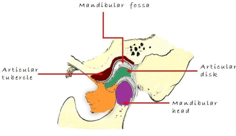
The temporomandibular joint (TMJ) can be viewed using various imaging techniques. Due to relatively low radiation doses and excellent spatial resolution, cone-beam computed tomography (CBCT) is being utilized more frequently in dental-maxillofacial imaging. For the diagnosis and treatment of TMJ disorders, an imaging examination is required. The osseous compartment is visualized using conventional CT, and CBCT and soft tissue imaging are extremely well appreciated on MRI. However, conventional TMJ imaging has its limitations due to its two-dimensional view and adjacent anatomical superimposition. TMJ imaging helps analyze the cortical and the bony compartment's trabaculae and assess the degree of skeletal abnormalities. TMJ imaging protocols are also used to evaluate treatment responses. CBCT is the three-dimensional imaging of the bony compartment and joint space and the morphology of the bone visualized by removing superimposition and distortion. Compared to multislice CT, CBCT produces high-resolution multiplanar images with a reduced dose of radiation. The role of CBCT imaging in determining the normal bony anatomy and pathological changes is appropriately delineated in this paper. This work will focus on the use of CBCT for the examination of TMJ in various patient categories, including those with osteoarthritis, remodeling, ankylosis, trauma, rheumatoid arthritis, synovial chondromatosis, and other intracapsular pathologies.
颞下颌关节(TMJ)可通过多种成像技术进行观察。由于辐射剂量相对较低且空间分辨率出色,锥形束计算机断层扫描(CBCT)在口腔颌面成像中的应用越来越频繁。对于颞下颌关节紊乱病的诊断和治疗,需要进行成像检查。使用传统CT可观察骨腔,而CBCT和软组织成像在MRI上的显示效果极佳。然而,传统的颞下颌关节成像由于其二维视图和相邻解剖结构的重叠而存在局限性。颞下颌关节成像有助于分析皮质骨和骨腔的骨小梁,并评估骨骼异常的程度。颞下颌关节成像方案也用于评估治疗反应。CBCT是对骨腔、关节间隙以及通过消除重叠和变形而可视化的骨形态进行三维成像。与多层CT相比,CBCT能产生高分辨率的多平面图像,且辐射剂量更低。本文对CBCT成像在确定正常骨解剖结构和病理变化方面的作用进行了恰当的描述。这项工作将聚焦于CBCT在各类患者中用于颞下颌关节检查的应用,包括患有骨关节炎、重塑、关节强直、创伤、类风湿关节炎、滑膜软骨瘤病以及其他关节内病变的患者。