Lasek Julia, Nurzynska Karolina, Piórkowski Adam, Strzelecki Michał, Obuchowicz Rafał
Faculty of Geology, Geophysics and Environmental Protection, AGH University of Krakow, 30-059 Krakow, Poland.
Department of Algorithmics and Software, Silesian University of Technology, 44-100 Gliwice, Poland.
Tomography. 2025 Feb 27;11(3):27. doi: 10.3390/tomography11030027.
Temporomandibular joint (TMJ) disorders are a significant cause of orofacial pain. Artificial intelligence (AI) has been successfully applied to other imaging modalities but remains underexplored in ultrasonographic evaluations of TMJ.
This study aimed to develop and validate an AI-driven method for the automatic and reproducible measurement of TMJ space width from ultrasonographic images.
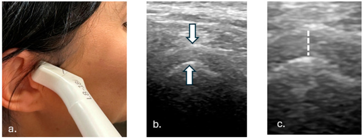
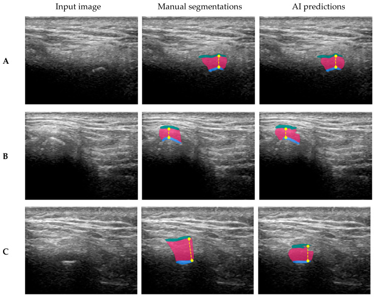
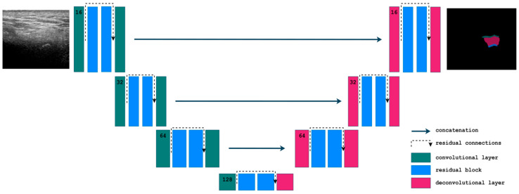
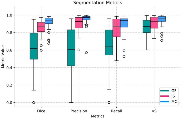
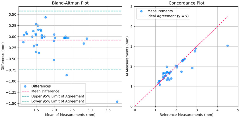
A total of 142 TMJ ultrasonographic images were segmented into three anatomical components: the mandibular condyle, joint space, and glenoid fossa. State-of-the-art architectures were tested, and the best-performing 2D Residual U-Net was trained and validated against expert annotations. The algorithm for joint space width measurement based on TMJ segmentation was proposed, calculating the vertical distance between the superior-most point of the mandibular condyle and its corresponding point on the glenoid fossa.
The segmentation model achieved high performance for the mandibular condyle (Dice: 0.91 ± 0.08) and joint space (Dice: 0.86 ± 0.09), with notably lower performance for the glenoid fossa (Dice: 0.60 ± 0.24), highlighting variability due to its complex geometry. The TMJ space width measurement algorithm demonstrated minimal bias, with a mean difference of 0.08 mm and a mean absolute error of 0.18 mm compared to reference measurements.
The model exhibited potential as a reliable tool for clinical use, demonstrating accuracy in TMJ ultrasonographic analysis. This study underscores the ability of AI-driven segmentation and measurement algorithms to bridge existing gaps in ultrasonographic imaging and lays the foundation for broader clinical applications.
颞下颌关节(TMJ)紊乱是口面部疼痛的一个重要原因。人工智能(AI)已成功应用于其他成像方式,但在TMJ的超声评估中仍未得到充分探索。
本研究旨在开发并验证一种由人工智能驱动的方法,用于从超声图像中自动且可重复地测量TMJ间隙宽度。
总共142张TMJ超声图像被分割为三个解剖成分:下颌髁突、关节间隙和关节盂。测试了最先进的架构,并针对专家标注对表现最佳的二维残差U-Net进行了训练和验证。提出了基于TMJ分割的关节间隙宽度测量算法,计算下颌髁突最上点与其在关节盂上对应点之间的垂直距离。
分割模型在下颌髁突(骰子系数:0.91±0.08)和关节间隙(骰子系数:0.86±0.09)方面表现出高性能,而在关节盂方面表现明显较低(骰子系数:0.60±0.24),突出了由于其复杂几何形状导致的变异性。TMJ间隙宽度测量算法显示出最小偏差,与参考测量相比,平均差异为0.08毫米,平均绝对误差为0.18毫米。
该模型展现出作为临床可靠工具的潜力,在TMJ超声分析中显示出准确性。本研究强调了人工智能驱动的分割和测量算法弥合超声成像现有差距的能力,并为更广泛的临床应用奠定了基础。