Murdoch Children's Research Institute, Melbourne, Victoria, Australia.
Royal Children's Hospital Melbourne, Melbourne, Victoria, Australia.
J Glob Health. 2022 Dec 23;12:10013. doi: 10.7189/jogh.12.10013.
Chest x-ray (CXR) is commonly used (when available) to support clinical management decisions for child pneumonia and provide a reference standard for diagnosis in research studies. However, its diagnostic and technical limitations for both purposes are well recognised. Recent evidence suggests that lung ultrasound (LUS) may have diagnostic utility in pneumonia. This systematic scoping review of research on the utility of CXR and LUS in the management of severe childhood pneumonia aims to inform pragmatic guidelines for low- and middle-income countries (LMICs) and identify gaps in knowledge.
We included peer-reviewed studies published between 2000 and 2020 in infants and children aged from one month to nine years, presenting with severe pneumonia. CXR studies were limited to those from LMICs, while LUS studies included any geographic region. LUS and CXR articles were mapped into the following themes: indications, role in diagnosis, role in management, impact on outcomes, and practical considerations for LMIC settings.
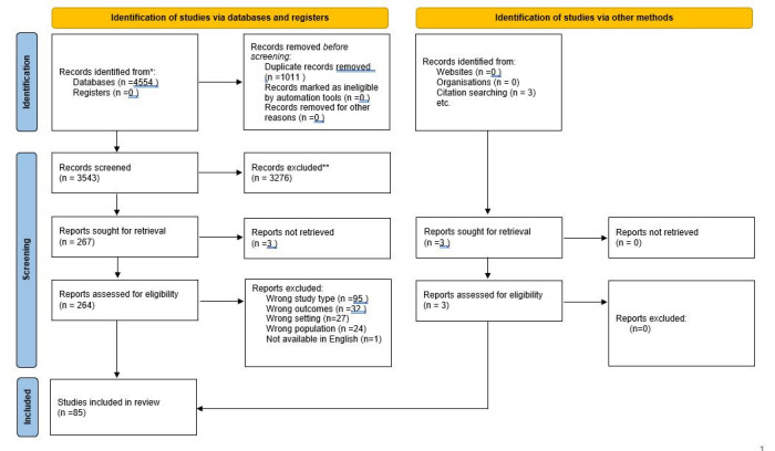
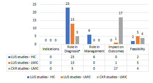
85 articles met all eligibility criteria, including 27 CXR studies and 58 LUS studies. CXR studies were primarily observational and examined associations between radiographic abnormalities and pneumonia aetiology or outcomes. The most consistent finding was an association between CXR consolidation and risk of mortality. Difficulty obtaining quality CXR images and inter-reader variability in interpretation were commonly reported challenges. Research evaluating indications for CXR, role in management, and impact on patient outcomes was very limited. LUS studies primarily focused on diagnostic accuracy. LUS had higher sensitivity for identification of consolidation than CXR. There are gaps in knowledge regarding diagnostic criteria, as well as the practical utility of LUS in the diagnosis and management of pneumonia. Most LUS studies were conducted in HIC settings with experienced operators; however, small feasibility studies indicate that good inter-operator reliability may be achieved by training of novice clinicians in LMIC settings.
The available evidence does not support the routine use of CXR or LUS as essential tools in the diagnosis and initial management of severe pneumonia. Further evaluation is required to determine the clinical utility and feasibility of both imaging modalities in low-resource settings.
胸部 X 光(CXR)常用于支持儿童肺炎的临床管理决策,并为研究中的诊断提供参考标准。然而,其在这两个目的下的诊断和技术限制是众所周知的。最近的证据表明,肺部超声(LUS)在肺炎的诊断中可能具有实用价值。本系统评价综述了 CXR 和 LUS 在管理严重儿童肺炎中的效用研究,旨在为中低收入国家(LMIC)提供实用指南,并确定知识空白。
我们纳入了 2000 年至 2020 年期间发表的、年龄在 1 个月至 9 岁的患有严重肺炎的婴儿和儿童的同行评审研究。CXR 研究仅限于来自 LMIC 的研究,而 LUS 研究包括任何地理区域。LUS 和 CXR 文章被映射到以下主题:适应症、诊断作用、管理作用、对结果的影响以及对 LMIC 环境的实际考虑。
85 篇文章符合所有入选标准,包括 27 篇 CXR 研究和 58 篇 LUS 研究。CXR 研究主要是观察性的,研究了放射异常与肺炎病因或结果之间的关联。最一致的发现是 CXR 实变与死亡率之间的关联。难以获得高质量的 CXR 图像和读者间解释的变异性是常见的报告挑战。评估 CXR 的适应症、管理作用和对患者结果的影响的研究非常有限。LUS 研究主要集中在诊断准确性上。与 CXR 相比,LUS 对实变的识别具有更高的敏感性。在诊断标准以及 LUS 在肺炎的诊断和管理中的实际应用方面存在知识空白。大多数 LUS 研究都是在 HIC 环境中由经验丰富的操作人员进行的;然而,一些小型可行性研究表明,通过在 LMIC 环境中培训新手临床医生,可以实现良好的操作者间可靠性。
现有证据不支持常规使用 CXR 或 LUS 作为严重肺炎诊断和初始管理的基本工具。需要进一步评估这两种成像方式在资源匮乏环境中的临床实用性和可行性。