School of Graduate Studies, Biomedical Sciences, Rutgers Biomedical and Health Sciences, Newark, New Jersey (Dr de Souza); Departments of Rehabilitation and Movement Sciences (Dr Esopenko) and (Drs Jia and Parrott), School of Health Professions, Rutgers Biomedical and Health Sciences, Newark, New Jersey; Department of Psychology and Neuroscience Center, Brigham Young University, Provo, Utah (Dr Merkley); Traumatic Brain Injury and Concussion Center, Department of Neurology, University of Utah School of Medicine, Salt Lake City (Drs Merkley, Dennis, Wilde, and Tate and Ms Velez); George E. Wahlen Department of Veterans Affairs Medical Center, Salt Lake City, Utah (Drs Dennis, Wilde, and Tate); Department of Psychology, Pennsylvania State University, University Park, and Social Life and Engineering Sciences Imaging Center, University Park, Pennsylvania (Dr Hillary); San Antonio VA Polytrauma Rehabilitation Center, San Antonio, Texas, and Departments of Rehabilitation Medicine and Psychiatry, UT Health San Antonio, San Antonio, Texas (Dr Cooper); TBI Center of Excellence at Joint Base San Antonio, Ft. Sam Houston, Texas AND Contractor, General Dynamics Information Technology, Silver Spring, Maryland (Dr Kennedy); Neurology Clinic, Wright Patterson Air Force Base, Wright Patterson AFB, Ohio (Dr Lewis); Alaska Radiology Associates, Anchorage, Alaska (Dr York); Michael E. DeBakey VA Medical Center, Houston, Texas (Dr Menefee); The Menninger Department of Psychiatry and Behavioral Sciences (Dr Menefee), Department of Neurology (Dr McCauley), H. Ben Taub Department of Physical Medicine and Rehabilitation (Drs McCauley and Wilde), and Department of Pediatrics (Dr McCauley), Baylor College of Medicine, Houston, Texas; and Brain Injury Rehabilitation Service, Department of Rehabilitation Medicine, Brooke Army Medical Center, Joint Base San Antonio, Fort Sam Houston, Texas (Dr Bowles). Drs de Souza and Esopenko are now at the Department of Rehabilitation and Human Performance, Icahn School of Medicine at Mount Sinai, New York, New York.
J Head Trauma Rehabil. 2023;38(4):E254-E266. doi: 10.1097/HTR.0000000000000848. Epub 2022 Dec 30.
Mild traumatic brain injury (mTBI) and posttraumatic stress disorder (PTSD) commonly occur among military Service Members and Veterans and have heterogenous, but also overlapping symptom presentations, which often complicate the diagnoses of underlying impairments and development of effective treatment plans. Thus, we sought to examine whether the combination of whole brain gray matter (GM) and white matter (WM) structural measures with neuropsychological performance can aid in the classification of military personnel with mTBI and PTSD.
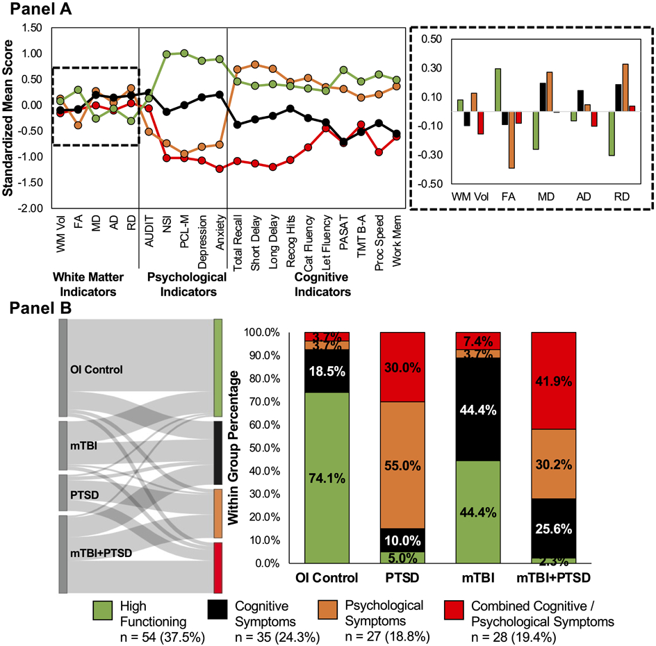
Active-Duty US Service Members ( n = 156; 87.8% male) with a history of mTBI, PTSD, combined mTBI+PTSD, or orthopedic injury completed a neuropsychological battery and T1- and diffusion-weighted structural neuroimaging. Cortical, subcortical, ventricular, and WM volumes and whole brain fractional anisotropy (FA), mean diffusivity (MD), radial diffusivity (RD), and axial diffusivity (AD) were calculated. Latent profile analyses were performed to determine how the GM and WM indicators, together with neuropsychological indicators, classified individuals.
For both GM and WM, respectively, a 4-profile model was the best fit. The GM model identified greater ventricular volumes in Service Members with cognitive symptoms, including those with a diagnosis of mTBI, either alone or with PTSD. The WM model identified reduced FA and elevated RD in those with psychological symptoms, including those with PTSD or mTBI and comorbid PTSD. However, contrary to expectation, a global neural signature unique to those with comorbid mTBI and PTSD was not identified.
The findings demonstrate that neuropsychological performance alone is more robust in differentiating Active-Duty Service Members with mTBI and PTSD, whereas global neuroimaging measures do not reliably differentiate between these groups.
轻度创伤性脑损伤(mTBI)和创伤后应激障碍(PTSD)在军人和退伍军人中很常见,它们具有异质的表现,也有重叠的症状,这往往使潜在损伤的诊断和有效治疗计划的制定变得复杂。因此,我们试图研究全脑灰质(GM)和白质(WM)结构测量与神经心理学表现的结合是否有助于对患有 mTBI 和 PTSD 的军人进行分类。
有 mTBI、PTSD、mTBI+PTSD 或骨科损伤病史的现役美国军人(n=156;87.8%为男性)完成了神经心理学测试和 T1 和弥散加权结构神经影像学检查。计算了皮质、皮质下、脑室和 WM 体积以及全脑各向异性分数(FA)、平均弥散系数(MD)、径向弥散系数(RD)和轴向弥散系数(AD)。进行潜在剖面分析,以确定 GM 和 WM 指标与神经心理学指标如何对个体进行分类。
对于 GM 和 WM,分别为 4 个模型的拟合度最好。GM 模型在有认知症状的军人中识别出更大的脑室体积,包括那些有 mTBI 诊断的军人,无论是单独患有还是同时患有 PTSD。WM 模型在有心理症状的军人中识别出 FA 降低和 RD 升高,包括那些患有 PTSD 或 mTBI 以及并发 PTSD 的军人。然而,与预期相反,并没有发现一种独特于并发 mTBI 和 PTSD 军人的全脑神经特征。
研究结果表明,神经心理学表现单独在区分患有 mTBI 和 PTSD 的现役军人方面更具优势,而全局神经影像学测量并不能可靠地区分这些群体。