• 文献检索
  • 文档翻译
  • 深度研究
  • 学术资讯
  • Suppr Zotero 插件Zotero 插件
  • 邀请有礼
  • 套餐&价格
  • 历史记录
应用&插件
Suppr Zotero 插件Zotero 插件浏览器插件Mac 客户端Windows 客户端微信小程序
定价
高级版会员购买积分包购买API积分包
服务
文献检索文档翻译深度研究API 文档MCP 服务
关于我们
关于 Suppr公司介绍联系我们用户协议隐私条款
关注我们

Suppr 超能文献

核心技术专利:CN118964589B侵权必究
粤ICP备2023148730 号-1Suppr @ 2026

文献检索

告别复杂PubMed语法,用中文像聊天一样搜索,搜遍4000万医学文献。AI智能推荐,让科研检索更轻松。

立即免费搜索

文件翻译

保留排版,准确专业,支持PDF/Word/PPT等文件格式,支持 12+语言互译。

免费翻译文档

深度研究

AI帮你快速写综述,25分钟生成高质量综述,智能提取关键信息,辅助科研写作。

立即免费体验

横断面肋下超声心动图:房室间隔缺损与短轴切面

Cross sectional subcostal echocardiography: atrioventricular septal defects and the short axis cut.

作者信息

Mortera C, Rissech M, Payola M, Miro C, Perich R

机构信息

Cardiologia Pediatrica, Hospital Infantil San Juan de Dios, Barcelona, Spain.

出版信息

Br Heart J. 1987 Sep;58(3):267-73. doi: 10.1136/hrt.58.3.267.

DOI:10.1136/hrt.58.3.267
PMID:3663428
原文链接:https://pmc.ncbi.nlm.nih.gov/articles/PMC1216448/
Abstract

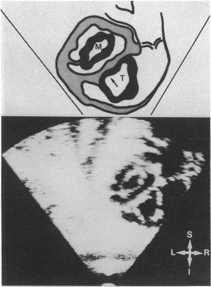
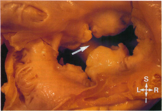
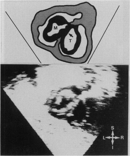
A short axis echocardiographic cut of the heart from the subcostal approach was used to study the atrioventricular junction in 47 infants and children with congenital heart disease and 20 with normal hearts. Examination of the diastolic openings of both atrioventricular valves was able to establish normal developments of the valves and annuli even when this was found in cases of complex congenital heart disease. In 30 patients with atrioventricular septal defects the technique distinguished between a partial defect (when the two atrioventricular valves were linked transseptally) and a complete defect (when there was only one atrioventricular valve). A range of atrioventricular attachments was seen in these patients. Short axis echocardiography from the subcostal approach reliably identifies different forms of atrioventricular septal defects by defining the anatomy of the atrioventricular valves during maximal diastolic expansion.

摘要

采用经肋下途径的心脏短轴超声心动图切面,对47例先天性心脏病患儿及20例心脏正常的婴幼儿的房室交界区进行研究。即使在复杂先天性心脏病病例中,对两个房室瓣舒张期开口的检查也能够确定瓣膜和瓣环的正常发育情况。在30例房室间隔缺损患者中,该技术能够区分部分缺损(两个房室瓣经间隔相连时)和完全缺损(只有一个房室瓣时)。在这些患者中观察到一系列房室连接情况。经肋下途径的短轴超声心动图通过在最大舒张期扩张时确定房室瓣的解剖结构,可靠地识别出不同形式的房室间隔缺损。

https://cdn.ncbi.nlm.nih.gov/pmc/blobs/81ec/1216448/38441ca8cdfe/brheartj00093-0095-a.jpg
https://cdn.ncbi.nlm.nih.gov/pmc/blobs/81ec/1216448/5b96af1d4c71/brheartj00093-0090-a.jpg
https://cdn.ncbi.nlm.nih.gov/pmc/blobs/81ec/1216448/50f01ccd5407/brheartj00093-0091-a.jpg
https://cdn.ncbi.nlm.nih.gov/pmc/blobs/81ec/1216448/64061fb2a6fe/brheartj00093-0092-a.jpg
https://cdn.ncbi.nlm.nih.gov/pmc/blobs/81ec/1216448/00ea1f4679e0/brheartj00093-0093-a.jpg
https://cdn.ncbi.nlm.nih.gov/pmc/blobs/81ec/1216448/541347e60d0d/brheartj00093-0094-a.jpg
https://cdn.ncbi.nlm.nih.gov/pmc/blobs/81ec/1216448/8e31086dba79/brheartj00093-0094-b.jpg
https://cdn.ncbi.nlm.nih.gov/pmc/blobs/81ec/1216448/38441ca8cdfe/brheartj00093-0095-a.jpg

相似文献

1
Cross sectional subcostal echocardiography: atrioventricular septal defects and the short axis cut.横断面肋下超声心动图:房室间隔缺损与短轴切面
Br Heart J. 1987 Sep;58(3):267-73. doi: 10.1136/hrt.58.3.267.
2
Cross-sectional echocardiographic assessment of conditions with atrioventricular valve leaflets attached to the atrial septum at the same level.经胸超声心动图对房室瓣小叶附着于同一水平房间隔的情况进行横断面评估。
Br Heart J. 1982 Oct;48(4):331-41. doi: 10.1136/hrt.48.4.331.
3
Mitral and tricuspid valve repair and growth in unbalanced atrial ventricular canal defects.二尖瓣和三尖瓣修复和增长在非均衡性房室管缺损中。
J Thorac Cardiovasc Surg. 2012 Apr;143(4 Suppl):S29-32. doi: 10.1016/j.jtcvs.2011.10.031. Epub 2011 Dec 6.
4
[Dysplasia of both the components of the atrioventricular valve in atrioventricular septal defects].
G Ital Cardiol. 1993 Aug;23(8):801-7.
5
Subxiphoid M-mode echocardiography in atrioventricular defects.
Pediatr Cardiol. 1982;3(2):119-25. doi: 10.1007/BF02312958.
6
[Quantitative evaluation of complete endocardial cushion defect using two-dimensional echocardiography].[二维超声心动图对完全性心内膜垫缺损的定量评估]
J Cardiol. 1987 Mar;17(1):169-78.
7
Cross-sectional echocardiography in the diagnosis of atrioventricular septal defect.经胸超声心动图在房室间隔缺损诊断中的应用
Int J Cardiol. 1990 Jul;28(1):19-23. doi: 10.1016/0167-5273(90)90004-o.
8
Differential and linear insertion of atrioventricular valves: a useful tool?房室瓣的差异和线性植入:一种有用的工具?
Ultrasound Obstet Gynecol. 2014 Nov;44(5):568-74. doi: 10.1002/uog.13326.
9
[Prenatal echocardiographic diagnosis of a case of atrioventricular septum defect].[一例房室间隔缺损的产前超声心动图诊断]
Rev Esp Cardiol. 1985 Jul-Aug;38(4):288-90.
10
Atrioventricular valve anular diameter: two-dimensional echocardiographic-autopsy correlation.
Am J Cardiol. 1984 Jun 1;53(11):1652-5. doi: 10.1016/0002-9149(84)90596-4.

引用本文的文献

1
Subcostal Echocardiographic Imaging in Neonatal and Pediatric Intensive Care.新生儿及儿科重症监护中的肋下超声心动图成像
Front Pediatr. 2021 Jun 24;9:471558. doi: 10.3389/fped.2021.471558. eCollection 2021.
2
Echocardiography in adult congenital heart disease.成人先天性心脏病的超声心动图检查
Heart. 1998 Nov;80 Suppl 1(Suppl 1):S12-26. doi: 10.1136/hrt.80.2008.12s.

本文引用的文献

1
Endocardial cushion defects; common atrio-ventricular canal and ostium primum.心内膜垫缺损;共同房室通道和原发孔。
Br Heart J. 1957 Jul;19(3):403-18. doi: 10.1136/hrt.19.3.403.
2
Surgical anatomy of the atrioventricular valve in the intermediate type of common atrioventricular orifice.共同房室口中间型房室瓣的手术解剖
J Thorac Cardiovasc Surg. 1980 Jun;79(6):884-9.
3
Sub-xiphoid echocardiography--a new approach to the diagnosis and differentiation of atrioventricular defects.剑突下超声心动图——一种诊断和鉴别房室缺损的新方法。
Eur Heart J. 1980 Feb;1(1):45-54. doi: 10.1093/oxfordjournals.eurheartj.a061094.
4
Subxiphoid M-mode echocardiography in atrioventricular defects.
Pediatr Cardiol. 1982;3(2):119-25. doi: 10.1007/BF02312958.
5
Assessment of atrioventricular septal defects by two dimensional echocardiography.二维超声心动图评估房室间隔缺损
Br Heart J. 1982 Feb;47(2):109-21. doi: 10.1136/hrt.47.2.109.
6
Internal cardiac crux: two-dimensional echocardiography of normal and congenitally abnormal hearts.心脏内部交叉点:正常及先天性异常心脏的二维超声心动图
Ultrasound Med Biol. 1984 Nov-Dec;10(6):735-45. doi: 10.1016/0301-5629(84)90234-5.
7
Anatomic observations on complete form of persistent common atrioventricular canal with special reference to atrioventricular valves.完全型永存共同房室通道的解剖学观察,特别涉及房室瓣
Mayo Clin Proc. 1966 May;41(5):296-308.
8
Echocardiographic features of endocardial cushion defects.心内膜垫缺损的超声心动图特征。
Circulation. 1974 Mar;49(3):418-22. doi: 10.1161/01.cir.49.3.418.
9
Multiple crystal echocardiographic evaluation of endocardial cushion defect.心内膜垫缺损的多晶体超声心动图评估
Circulation. 1974 Jul;50(1):25-32. doi: 10.1161/01.cir.50.1.25.
10
The spectrum of common atrioventricular orifice (canal).常见房室口(管)的频谱。
Am Heart J. 1973 Oct;86(4):553-61. doi: 10.1016/0002-8703(73)90148-8.