• 文献检索
  • 文档翻译
  • 深度研究
  • 学术资讯
  • Suppr Zotero 插件Zotero 插件
  • 邀请有礼
  • 套餐&价格
  • 历史记录
应用&插件
Suppr Zotero 插件Zotero 插件浏览器插件Mac 客户端Windows 客户端微信小程序
定价
高级版会员购买积分包购买API积分包
服务
文献检索文档翻译深度研究API 文档MCP 服务
关于我们
关于 Suppr公司介绍联系我们用户协议隐私条款
关注我们

Suppr 超能文献

核心技术专利:CN118964589B侵权必究
粤ICP备2023148730 号-1Suppr @ 2026

文献检索

告别复杂PubMed语法,用中文像聊天一样搜索,搜遍4000万医学文献。AI智能推荐,让科研检索更轻松。

立即免费搜索

文件翻译

保留排版,准确专业,支持PDF/Word/PPT等文件格式,支持 12+语言互译。

免费翻译文档

深度研究

AI帮你快速写综述,25分钟生成高质量综述,智能提取关键信息,辅助科研写作。

立即免费体验

基于动态对比增强超声定量分析预测胰腺神经内分泌肿瘤的病理分级

Prediction of Pathological Grades of Pancreatic Neuroendocrine Tumors Based on Dynamic Contrast-Enhanced Ultrasound Quantitative Analysis.

作者信息

Yang Dao-Hui, Cheng Juan, Tian Xiao-Fan, Zhang Qi, Yu Ling-Yun, Qiu Yi-Jie, Lu Xiu-Yun, Lou Wen-Hui, Dong Yi, Wang Wen-Ping

机构信息

Department of Ultrasound, Zhongshan Hospital, Fudan University (Xiamen Branch), Xiamen 361006, China.

Department of Ultrasound, Xinhua Hospital Affiliated to Shanghai Jiaotong University School of Medicine, Shanghai 200092, China.

出版信息

Diagnostics (Basel). 2023 Jan 9;13(2):238. doi: 10.3390/diagnostics13020238.

DOI:10.3390/diagnostics13020238
PMID:36673048
原文链接:https://pmc.ncbi.nlm.nih.gov/articles/PMC9858178/
Abstract

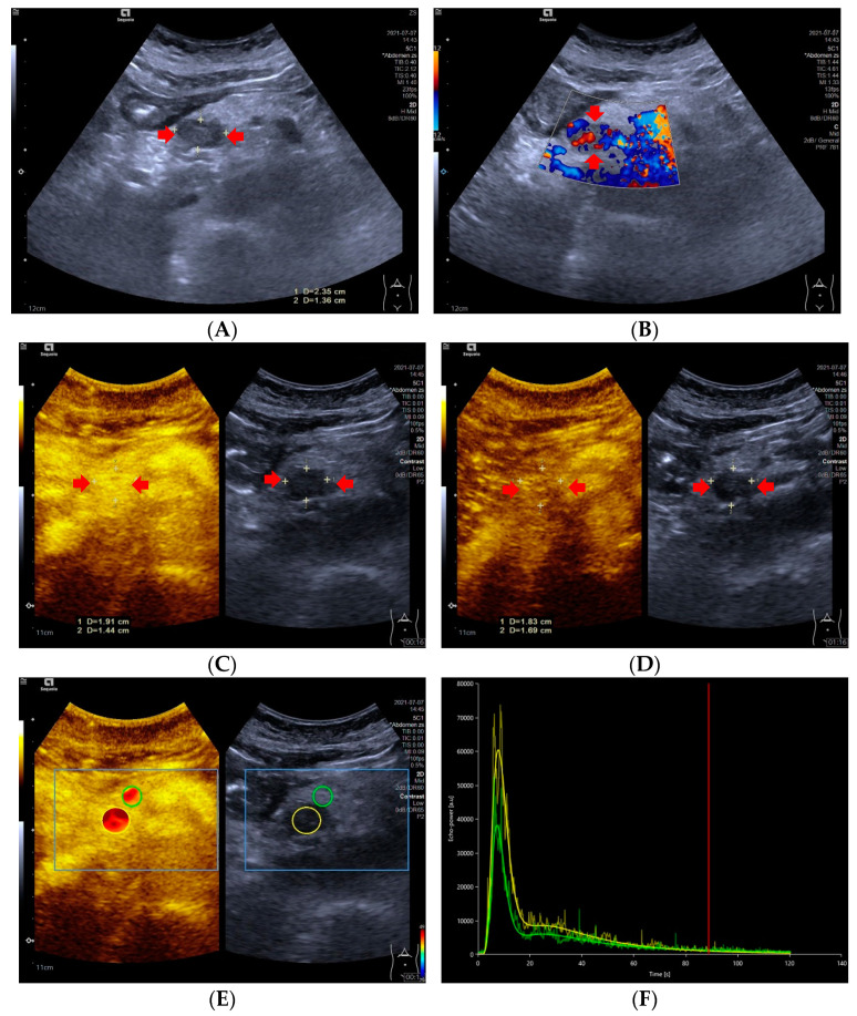
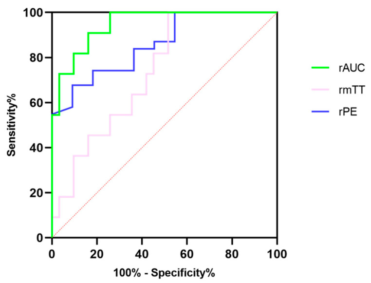
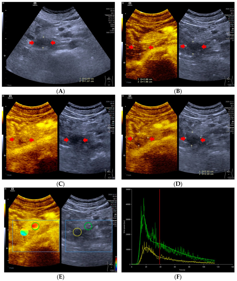
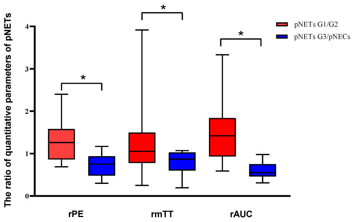
Objective: To investigate whether the dynamic contrast-enhanced ultrasound (DCE-US) analysis and quantitative parameters could be helpful for predicting histopathologic grades of pancreatic neuroendocrine tumors (pNETs). Methods: This retrospective study conducted a comprehensive review of the CEUS database between March 2017 and November 2021 in Zhongshan Hospital, Fudan University. Ultrasound examinations were performed by an ACUSON Sequioa unit equipped with a 3.5 MHz 6C−1 convex array transducer, and an ACUSON OXANA2 unit equipped with a 3.5 MHz 5C−1 convex array transducer. SonoVue® (Bracco Inc., Milan, Italy) was used for all CEUS examinations. Time intensity curves (TICs) and quantitative parameters of DCE-US were created by Vuebox® software (Bracco, Italy). Inclusion criteria were: patients with histopathologically proved pNETs, patients who underwent pancreatic B-mode ultrasounds (BMUS) and CEUS scans one week before surgery or biopsy and had DCE-US imaging documented for more than 2 min, patients with solid or predominantly solid lesions and patients with definite diagnosis of histopathological grades of pNETs. Based on their prognosis, patients were categorized into two groups: pNETs G1/G2 group and pNETs G3/pNECs group. Results: A total of 42 patients who underwent surgery (n = 38) or biopsy (n = 4) and had histopathologically confirmed pNETs were included. According to the WHO 2019 criteria, all pNETs were classified into grade 1 (G1, n = 10), grade 2 (G2, n = 21), or grade 3 (G3)/pancreatic neuroendocrine carcinomas (pNECs) (n = 11), based on the Ki−67 proliferation index and the mitotic activity. The majority of the TICs (27/31) of pNETs G1/G2 were above or equal to those of pancreatic parenchyma in the arterial phase, but most (7/11) pNETs G3/pNECs had TICs below those of pancreatic parenchyma from arterial phase to late phase (p < 0.05). Among all the CEUS quantitative parameters of DCE-US, values of relative rise time (rPE), relative mean transit time (rmTT) and relative area under the curve (rAUC) were significantly higher in pNETs G1/G2 group than those in pNETs G3/pNECs group (p < 0.05). Taking an rPE below 1.09 as the optimal cut-off value, the sensitivity, specificity and accuracy for prediction of pNETs G3/pNECs from G1/G2 were 90.91% [58.70% to 99.80%], 67.64% [48.61% to 83.32%] and 85.78% [74.14% to 97.42%], respectively. Taking rAUC below 0.855 as the optimal cut-off value, the sensitivity, specificity and accuracy for prediction of pNETs G3/pNECs from G1/G2 were 90.91% [66.26% to 99.53%], 83.87% [67.37% to 92.91%] and 94.72% [88.30% to 100.00%], respectively. Conclusions: Dynamic contrast-enhanced ultrasound analysis might be helpful for predicting the pathological grades of pNETs. Among all quantitative parameters, rPE, rmTT and rAUC are potentially useful parameters for predicting G3/pNECs with aggressive behavior.

摘要

目的

探讨动态对比增强超声(DCE-US)分析及定量参数是否有助于预测胰腺神经内分泌肿瘤(pNETs)的组织病理学分级。方法:本回顾性研究对2017年3月至2021年11月复旦大学附属中山医院的CEUS数据库进行了全面回顾。超声检查采用配备3.5 MHz 6C−1凸阵探头的ACUSON Sequioa超声仪和配备3.5 MHz 5C−1凸阵探头的ACUSON OXANA2超声仪。所有CEUS检查均使用声诺维®(意大利米兰百胜公司)。DCE-US的时间强度曲线(TICs)和定量参数由Vuebox®软件(意大利百胜)生成。纳入标准为:组织病理学证实为pNETs的患者;术前或活检前一周接受过胰腺B超(BMUS)和CEUS扫描且DCE-US成像记录超过2分钟的患者;实性或主要为实性病变的患者;pNETs组织病理学分级明确诊断的患者。根据预后情况,将患者分为两组:pNETs G1/G2组和pNETs G3/pNECs组。结果:共纳入42例接受手术(n = 38)或活检(n = 4)且组织病理学确诊为pNETs的患者。根据世界卫生组织2019年标准,基于Ki−67增殖指数和有丝分裂活性,所有pNETs分为1级(G1,n = 10)、2级(G2,n = 21)或3级(G3)/胰腺神经内分泌癌(pNECs)(n = 11)。pNETs G1/G2的大多数TICs(27/31)在动脉期高于或等于胰腺实质,但大多数(7/11)pNETs G3/pNECs从动脉期到晚期的TICs低于胰腺实质(p < 0.05)。在DCE-US的所有CEUS定量参数中,pNETs G1/G2组的相对上升时间(rPE)、相对平均通过时间(rmTT)和相对曲线下面积(rAUC)值显著高于pNETs G3/pNECs组(p < 0.05)。以rPE低于1.09作为最佳截断值,从G1/G2预测pNETs G3/pNECs的敏感性、特异性和准确性分别为90.91%[58.70%至99.80%]、67.64%[48.61%至83.32%]和85.78%[74.14%至97.42%]。以rAUC低于0.855作为最佳截断值,从G1/G2预测pNETs G3/pNECs的敏感性、特异性和准确性分别为90.91%[66.26%至99.53%]、83.87%[67.37%至92.91%]和94.72%[88.30%至100.00%]。结论:动态对比增强超声分析可能有助于预测pNETs的病理分级。在所有定量参数中

https://cdn.ncbi.nlm.nih.gov/pmc/blobs/6107/9858178/491c473895d5/diagnostics-13-00238-g004.jpg
https://cdn.ncbi.nlm.nih.gov/pmc/blobs/6107/9858178/2d6e3c9dd6dd/diagnostics-13-00238-g001.jpg
https://cdn.ncbi.nlm.nih.gov/pmc/blobs/6107/9858178/0225a255261f/diagnostics-13-00238-g002.jpg
https://cdn.ncbi.nlm.nih.gov/pmc/blobs/6107/9858178/604a14d8f706/diagnostics-13-00238-g003.jpg
https://cdn.ncbi.nlm.nih.gov/pmc/blobs/6107/9858178/491c473895d5/diagnostics-13-00238-g004.jpg

相似文献

1
Prediction of Pathological Grades of Pancreatic Neuroendocrine Tumors Based on Dynamic Contrast-Enhanced Ultrasound Quantitative Analysis.基于动态对比增强超声定量分析预测胰腺神经内分泌肿瘤的病理分级
Diagnostics (Basel). 2023 Jan 9;13(2):238. doi: 10.3390/diagnostics13020238.
2
Early Assessment of Chemoradiotherapy Response for Locally Advanced Pancreatic Ductal Adenocarcinoma by Dynamic Contrast-Enhanced Ultrasound.动态对比增强超声对局部晚期胰腺导管腺癌放化疗反应的早期评估
Diagnostics (Basel). 2022 Nov 2;12(11):2662. doi: 10.3390/diagnostics12112662.
3
Contrast-enhanced ultrasound (CEUS) and shear wave elastography (SWE) features for characterizing serous microcystic adenomas (SMAs): In comparison to pancreatic neuroendocrine tumors (pNETs).用于表征浆液性微囊性腺瘤(SMA)的超声造影(CEUS)和剪切波弹性成像(SWE)特征:与胰腺神经内分泌肿瘤(pNET)相比。
Heliyon. 2024 Feb 1;10(3):e25185. doi: 10.1016/j.heliyon.2024.e25185. eCollection 2024 Feb 15.
4
Pancreatic neuroendocrine tumor: prediction of tumor grades by radiomics models based on ultrasound images.胰腺神经内分泌肿瘤:基于超声图像的放射组学模型预测肿瘤分级。
Br J Radiol. 2023 Sep;96(1149):20220783. doi: 10.1259/bjr.20220783. Epub 2023 Jul 26.
5
Applications of Dynamic Contrast-Enhanced Ultrasound in Differential Diagnosis of Hepatocellular Carcinoma and Intrahepatic Cholangiocarcinoma in Non-cirrhotic Liver.动态对比增强超声在非肝硬化肝脏肝细胞癌和肝内胆管细胞癌鉴别诊断中的应用。
Ultrasound Med Biol. 2023 Aug;49(8):1780-1788. doi: 10.1016/j.ultrasmedbio.2023.03.026. Epub 2023 May 6.
6
Clinical relevance of different WHO grade 3 pancreatic neuroendocrine neoplasms based on morphology.基于形态学的不同WHO 3级胰腺神经内分泌肿瘤的临床相关性
Endocr Connect. 2018 Feb;7(2):355-363. doi: 10.1530/EC-17-0388.
7
VueBox® perfusion analysis of dynamic contrast enhanced ultrasound provides added value in the diagnosis of small thyroid nodules.VueBox® 灌注分析在诊断甲状腺小结节中的应用
Clin Hemorheol Microcirc. 2023;83(4):409-420. doi: 10.3233/CH-221681.
8
Application of dynamic contrast enhanced ultrasound in distinguishing focal-type autoimmune pancreatitis from pancreatic ductal adenocarcinoma.动态对比增强超声在鉴别局灶型自身免疫性胰腺炎与胰腺导管腺癌中的应用
Clin Hemorheol Microcirc. 2022;81(2):149-161. doi: 10.3233/CH-221390.
9
Quantitative analysis of enhanced CT in differentiating well-differentiated pancreatic neuroendocrine tumors and poorly differentiated neuroendocrine carcinomas.增强 CT 定量分析在鉴别高分化胰腺神经内分泌肿瘤和低分化神经内分泌癌中的应用。
Eur Radiol. 2022 Dec;32(12):8317-8325. doi: 10.1007/s00330-022-08891-4. Epub 2022 Jun 27.
10
Application of Dynamic Contrast-Enhanced Ultrasound in Evaluation the Activity of Crohn's Disease.动态对比增强超声在评估克罗恩病活动度中的应用
Diagnostics (Basel). 2024 Mar 22;14(7):672. doi: 10.3390/diagnostics14070672.

引用本文的文献

1
Contrast-Enhanced Harmonic Endoscopic Ultrasonography for Prediction of Aggressiveness and Treatment Response in Patients with Pancreatic Lesions.对比增强谐波内镜超声检查对胰腺病变患者侵袭性及治疗反应的预测作用
Cancers (Basel). 2025 Aug 1;17(15):2545. doi: 10.3390/cancers17152545.
2
Role of B-Mode and Contrast-Enhanced Ultrasound in the Diagnostic Workflow of Gastro-Entero-Pancreatic Neuroendocrine Tumors (GEP-NETs).B超及超声造影在胃肠胰神经内分泌肿瘤(GEP-NETs)诊断流程中的作用
Cancers (Basel). 2025 Jun 4;17(11):1879. doi: 10.3390/cancers17111879.
3
Endoscopic ultrasonography-based intratumoral and peritumoral machine learning ultrasomics model for predicting the pathological grading of pancreatic neuroendocrine tumors.

本文引用的文献

1
Preoperative assessment of microvessel density in nonfunctioning pancreatic neuroendocrine tumors (NF-PanNETs).非功能性胰腺神经内分泌肿瘤 (NF-PanNETs) 微血管密度的术前评估。
Surgery. 2022 Oct;172(4):1236-1244. doi: 10.1016/j.surg.2022.06.017. Epub 2022 Aug 8.
2
Reliability of grading preoperative pancreatic neuroendocrine tumors on EUS specimens: a systematic review with meta-analysis of aggregate and individual data.术前超声内镜标本上胰腺神经内分泌肿瘤分级的可靠性:汇总和个体数据的系统评价和荟萃分析。
Gastrointest Endosc. 2022 Dec;96(6):898-908.e23. doi: 10.1016/j.gie.2022.07.014. Epub 2022 Jul 19.
3
Trends in Incidence and Survival of Patients with Pancreatic Neuroendocrine Neoplasm, 1987-2016.
基于内镜超声的肿瘤内和肿瘤周围机器学习超声组学模型用于预测胰腺神经内分泌肿瘤的病理分级
BMC Med Imaging. 2025 Jan 18;25(1):22. doi: 10.1186/s12880-025-01555-x.
4
Application of Dynamic Contrast-Enhanced Ultrasound in Evaluation the Activity of Crohn's Disease.动态对比增强超声在评估克罗恩病活动度中的应用
Diagnostics (Basel). 2024 Mar 22;14(7):672. doi: 10.3390/diagnostics14070672.
5
Dynamic contrast enhanced ultrasound in gastrointestinal diseases: A current trend or an indispensable tool?动态对比增强超声在胃肠道疾病中的应用:当前趋势还是不可或缺的工具?
World J Gastroenterol. 2023 Jul 7;29(25):4021-4035. doi: 10.3748/wjg.v29.i25.4021.
1987 - 2016年胰腺神经内分泌肿瘤患者的发病率及生存率趋势
J Oncol. 2021 Dec 22;2021:4302675. doi: 10.1155/2021/4302675. eCollection 2021.
4
Incidence, prevalence, and survival trends for neuroendocrine neoplasms in Victoria, Australia, from 1982 to 2019: Based on site, grade, and region.澳大利亚维多利亚州 1982 年至 2019 年神经内分泌肿瘤的发病率、患病率和生存趋势:基于部位、分级和区域。
Asia Pac J Clin Oncol. 2022 Oct;18(5):e306-e317. doi: 10.1111/ajco.13671. Epub 2021 Nov 24.
5
Incidental nonfunctioning pancreatic neuroendocrine tumors: Contrast enhanced ultrasound features in diagnosis1.偶然发现的无功能性胰腺神经内分泌肿瘤:超声造影在诊断中的特征 1 。
Clin Hemorheol Microcirc. 2022;80(4):343-352. doi: 10.3233/CH-211269.
6
Neuroendocrine and Adrenal Tumors, Version 2.2021, NCCN Clinical Practice Guidelines in Oncology.神经内分泌和肾上腺肿瘤,第2.2021版,美国国立综合癌症网络(NCCN)肿瘤学临床实践指南
J Natl Compr Canc Netw. 2021 Jul 28;19(7):839-868. doi: 10.6004/jnccn.2021.0032.
7
Contrast-Enhanced Ultrasonography Findings Correlate with Pathologic Grades of Pancreatic Neuroendocrine Tumors.超声造影表现与胰腺神经内分泌肿瘤的病理分级相关。
Ultrasound Med Biol. 2021 Aug;47(8):2097-2106. doi: 10.1016/j.ultrasmedbio.2021.02.013. Epub 2021 Apr 29.
8
Contrast enhanced ultrasound quantitative parameters for assessing neoadjuvant chemotherapy response in patients with locally advanced breast cancer.对比增强超声定量参数用于评估局部晚期乳腺癌患者新辅助化疗反应。
Br J Radiol. 2021 May 1;94(1121):20201160. doi: 10.1259/bjr.20201160. Epub 2021 Apr 16.
9
Contrast-enhanced harmonic endoscopic ultrasound-guided fine-needle aspiration versus standard fine-needle aspiration in pancreatic masses: a meta-analysis.对比增强谐波内镜超声引导下细针穿刺与标准细针穿刺在胰腺肿块中的应用:一项荟萃分析。
Expert Rev Gastroenterol Hepatol. 2021 Jul;15(7):821-828. doi: 10.1080/17474124.2021.1880893. Epub 2021 Jan 26.
10
Potential application of dynamic contrast enhanced ultrasound in predicting microvascular invasion of hepatocellular carcinoma.动态对比增强超声在预测肝细胞癌微血管侵犯中的应用潜力。
Clin Hemorheol Microcirc. 2021;77(4):461-469. doi: 10.3233/CH-201085.