Department of Neurosurgery, University of Iowa Hospitals & Clinics and Stead Family Children's Hospital, Iowa City, IA, USA.
Department of Neurosurgery, University of Iowa Hospitals & Clinics, 200 Hawkins Drive, 1824 JPP, Iowa City, IA, 52242, USA.
J Med Case Rep. 2023 Jan 23;17(1):22. doi: 10.1186/s13256-023-03759-7.
Filar cysts are frequently found on neonatal ultrasound and are physiologically involuting structures with natural resolution. Hence, there has been no previous histologic correlation. Ventriculus terminalis is a focal central canal dilation in the conus medullaris and usually not clinically significant. Extra-axial cyst at the conus-filum junction connected to ventriculus terminalis is extremely rare, especially when associated with tethered lipomatous filum terminale and with progressive cyst enlargement.
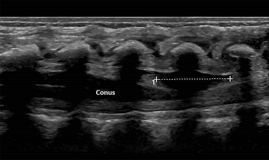
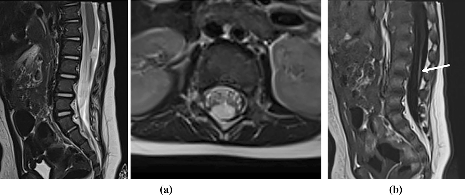
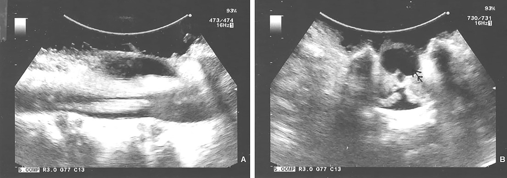
A Caucasian female neonate with abnormal gluteal cleft had ventriculus terminalis cyst with an extra-axial cyst at the conus-filar junction and taut lipomatous filum on ultrasound examination and magnetic resonance imaging. This persisted at 6-month follow up imaging. In light of the nonresolving extra-axial mass and thick taut lipomatous filum, the child underwent L1-L3 osteoplastic laminectomies. The extra-axial cyst expanded after bony decompression and furthermore on dural opening; visualized on ultrasound. It communicated with the central canal and was documented with intraoperative photomicrographs. It was excised and filum sectioned. Histological immunostaining of the cyst wall showed neuroglial and axonal elements. The child did well without deficits at 4-year follow up with normal urodynamics.
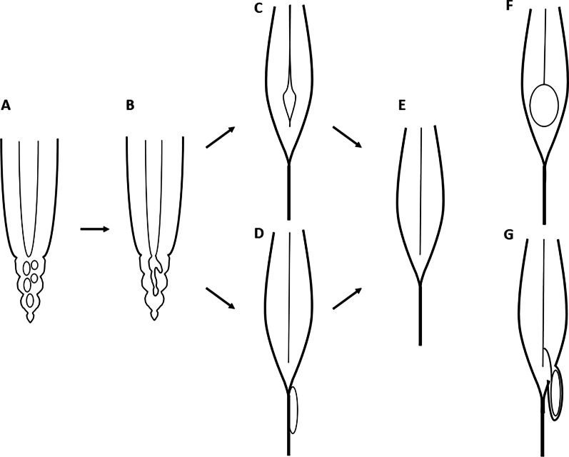
Progression dilation of ventriculus terminalis and extra-axial conofilar cyst with tethered lipomatous filum will likely progress to clinical significance and require surgical intervention. The embryologic basis for this pathology is discussed, with literature review.
新生儿超声检查常发现皮样囊肿,皮样囊肿是生理性退化结构,具有自然消退的特点。因此,以前没有进行过组织学相关性研究。终室是脊髓圆锥中央管的局灶性扩张,通常不具有临床意义。终丝圆锥-filum 交界处的硬膜外囊肿与终室相连非常罕见,尤其是当伴有脂肪性终丝马尾拴系和囊肿逐渐增大时。
一名高加索女性新生儿臀裂异常,超声和磁共振成像检查发现终室囊肿伴终丝圆锥-filum 交界处硬膜外囊肿和紧张性脂肪性终丝马尾。在 6 个月的随访成像中,这一情况仍存在。鉴于非消退性硬膜外肿块和紧张性肥厚性脂肪性终丝马尾,患儿接受了 L1-L3 骨成形椎板切除术。骨减压后和硬脑膜切开后,硬膜外囊肿扩大;在超声下可见。它与中央管相通,并通过术中照片记录下来。囊肿被切除,终丝被切断。囊肿壁的组织学免疫染色显示神经胶质和轴突成分。患儿在 4 年的随访中表现良好,无功能障碍,尿动力学正常。
终室扩张和硬膜外圆锥-filum 囊肿伴脂肪性终丝马尾拴系,可能会进展为具有临床意义的疾病,需要手术干预。讨论了这种病理学的胚胎学基础,并进行了文献复习。