Tang Xiaolong, Sui Xue, Liu Yongshuo
Department of Clinical Laboratory Diagnostics, Binzhou Medical University, Binzhou, Shandong 256603, China.
Department of Clinical Laboratory, Binzhou Medical University Hospital, Binzhou, Shandong 256603, China.
Heliyon. 2023 Jan 7;9(1):e12873. doi: 10.1016/j.heliyon.2023.e12873. eCollection 2023 Jan.
PTPN2, a member of the non-receptor protein tyrosine phosphatases family, holds a crucial role in tumorigenesis and cancer immunotherapy. However, most studies on the role of PTPN2 in cancer are limited to specific cancer types. Therefore, this study aimed to investigate the prognostic significance of PTPN2 in human cancers and its function in the tumor microenvironment.
To shed light on this matter, we investigated the expression level, prognostic value, genomic alterations, molecular function, immune function, and immunotherapeutic predictive ability of PTPN2 in human cancers using the TCGA, GTEx, CGGA, GEO, cBioPortal, STRING, TISCH, TIMER2.0, ESTIMATE, and TIDE databases. Furthermore, the CCK-8 assay was utilized to detect the effect of PTPN2 on cell proliferation. Cell immunofluorescence analysis was performed to probe the cellular localization of PTPN2. Western blot was applied to examine the molecular targets downstream of PTPN2. Finally, a Nomogram model was constructed using the TCGA-LGG cohort and evaluated with calibration curves and time-dependent ROCs.
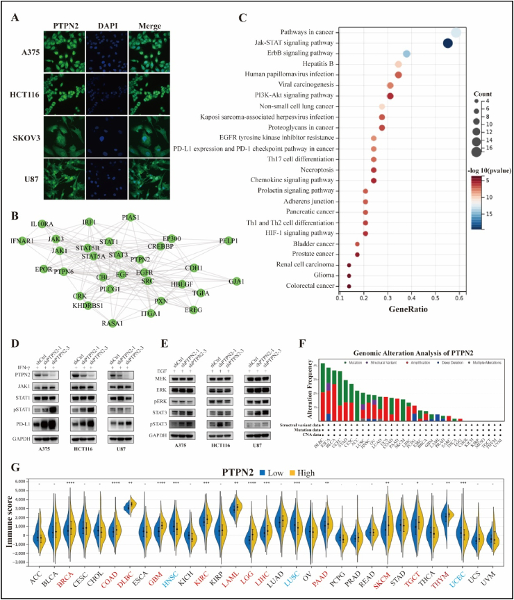
PTPN2 was highly expressed in most cancers and was linked to poor prognosis in ACC, GBM, LGG, KICH, and PAAD, while the opposite was true in OV, SKCM, and THYM. PTPN2 knockdown promoted the proliferation of melanoma cells, while significantly inhibiting proliferation in colon cancer and glioblastoma cells. In addition, TC-PTP, encoded by the PTPN2 gene, was primarily localized in the nucleus and cytoplasm and could negatively regulate the JAK/STAT and MEK/ERK pathways. Strikingly, PTPN2 knockdown significantly enhanced the abundance of PD-L1. PTPN2 was abundantly expressed in Mono/Macro cells and positively correlated with multiple immune infiltrating cells, especially CD8 T cells. Notably, DLBC, LAML, OV, and TGCT patients in the PTPN2-high group responded better to immunotherapy, while the opposite was true in ESCA, KIRC, KIRP, LIHC, and THCA. Finally, the construction of a Nomogram model on LGG exhibited a high prediction accuracy.
Immune checkpoint PTPN2 is a powerful biomarker for predicting prognosis and the efficacy of immunotherapy in cancers. Mechanistically, PTPN2 negatively regulates the JAK/STAT and MEK/ERK pathways and the abundance of PD-L1.
PTPN2是非受体蛋白酪氨酸磷酸酶家族的成员,在肿瘤发生和癌症免疫治疗中起着关键作用。然而,大多数关于PTPN2在癌症中作用的研究仅限于特定的癌症类型。因此,本研究旨在探讨PTPN2在人类癌症中的预后意义及其在肿瘤微环境中的功能。
为阐明这一问题,我们使用TCGA、GTEx、CGGA、GEO、cBioPortal、STRING、TISCH、TIMER2.0、ESTIMATE和TIDE数据库,研究了PTPN2在人类癌症中的表达水平、预后价值、基因组改变、分子功能、免疫功能和免疫治疗预测能力。此外,采用CCK-8试验检测PTPN2对细胞增殖的影响。进行细胞免疫荧光分析以探究PTPN2的细胞定位。应用蛋白质免疫印迹法检测PTPN2下游的分子靶点。最后,使用TCGA-LGG队列构建列线图模型,并通过校准曲线和时间依赖性ROC进行评估。
PTPN2在大多数癌症中高表达,与ACC、GBM、LGG、KICH和PAAD的不良预后相关,而在OV、SKCM和THYM中情况则相反。敲低PTPN2可促进黑色素瘤细胞增殖,同时显著抑制结肠癌细胞和胶质母细胞瘤细胞的增殖。此外,PTPN2基因编码的TC-PTP主要定位于细胞核和细胞质,可负调控JAK/STAT和MEK/ERK信号通路。引人注目的是,敲低PTPN2可显著提高PD-L1的丰度。PTPN2在单核/巨噬细胞中大量表达,与多种免疫浸润细胞呈正相关,尤其是CD8 T细胞。值得注意的是,PTPN2高表达组中的DLBC、LAML、OV和TGCT患者对免疫治疗反应更好,而在ESCA、KIRC、KIRP、LIHC和THCA中情况则相反。最后,基于LGG构建的列线图模型显示出较高的预测准确性。
免疫检查点PTPN2是预测癌症预后和免疫治疗疗效的有力生物标志物。机制上,PTPN2负调控JAK/STAT和MEK/ERK信号通路以及PD-L1的丰度。