Lucas Alfredo, Mouchtaris Sofia, Cornblath Eli J, Sinha Nishant, Caciagli Lorenzo, Hadar Peter, Gugger James J, Das Sandhitsu, Stein Joel M, Davis Kathryn A
Perelman School of Medicine, University of Pennsylvania.
Department of Bioengineering, University of Pennsylvania.
medRxiv. 2023 Jan 9:2023.01.08.23284313. doi: 10.1101/2023.01.08.23284313.
Functional gradients have been used to study differences in connectivity between healthy and diseased brain states, however this work has largely focused on the cortex. Because the subcortex plays a key role in seizure initiation in temporal lobe epilepsy (TLE), subcortical functional-connectivity gradients may help further elucidate differences between healthy brains and TLE, as well as differences between left (L)-TLE and right (R)-TLE.
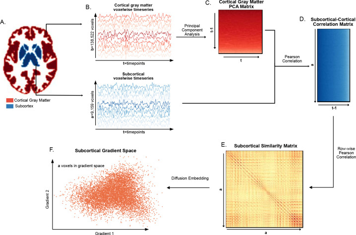
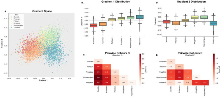
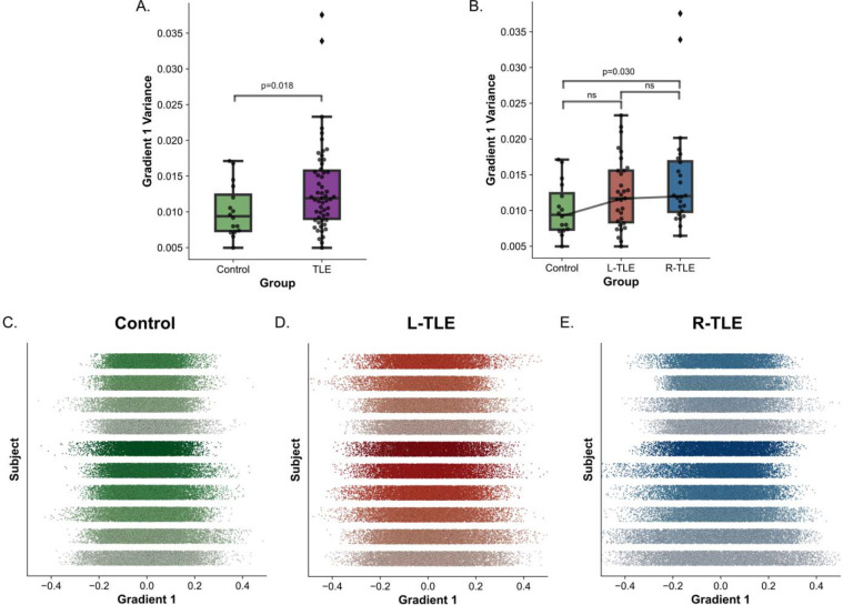
In this work, we calculated subcortical functional-connectivity gradients (SFGs) from resting-state functional MRI (rs-fMRI) by measuring the similarity in connectivity profiles of subcortical voxels to cortical gray matter voxels. We performed this analysis in 23 R-TLE patients and 32 L-TLE patients (who were otherwise matched for age, gender, disease specific characteristics, and other clinical variables), and 16 controls. To measure differences in SFGs between L-TLE and R-TLE, we quantified deviations in the average functional gradient distributions, as well as their variance, across subcortical structures.
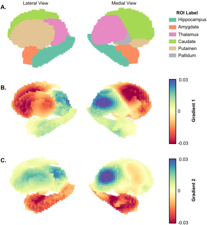
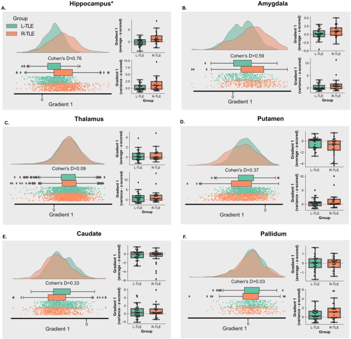
We found an expansion, measured by increased variance, in the principal SFG of TLE relative to controls. When comparing the gradient across subcortical structures between L-TLE and R-TLE, we found that abnormalities in the ipsilateral hippocampal gradient distributions were significantly different between L-TLE and R-TLE.
Our results suggest that expansion of the SFG is characteristic of TLE. Subcortical functional gradient differences exist between left and right TLE and are driven by connectivity changes in the hippocampus ipsilateral to the seizure onset zone.
功能梯度已被用于研究健康与患病脑状态之间的连接差异,然而这项工作主要集中在皮层。由于皮层下结构在颞叶癫痫(TLE)的癫痫发作起始中起关键作用,皮层下功能连接梯度可能有助于进一步阐明健康脑与TLE之间的差异,以及左侧(L)-TLE和右侧(R)-TLE之间的差异。
在本研究中,我们通过测量皮层下体素与皮层灰质体素连接图谱的相似性,从静息态功能磁共振成像(rs-fMRI)计算皮层下功能连接梯度(SFG)。我们对23例R-TLE患者、32例L-TLE患者(在年龄、性别、疾病特异性特征和其他临床变量方面进行了匹配)以及16名对照进行了此分析。为了测量L-TLE和R-TLE之间SFG的差异,我们量化了皮层下结构中平均功能梯度分布及其方差的偏差。
我们发现,相对于对照组,TLE的主要SFG通过方差增加来衡量出现了扩展。当比较L-TLE和R-TLE之间皮层下结构的梯度时,我们发现L-TLE和R-TLE之间同侧海马梯度分布的异常存在显著差异。
我们的结果表明,SFG的扩展是TLE的特征。左右TLE之间存在皮层下功能梯度差异,并且由癫痫发作起始区同侧海马的连接变化所驱动。