Edlin Joy, Nowell Justin, Arthurs Christopher, Figueroa Alberto, Jahangiri Marjan
Department of Cardiothoracic Surgery, St George's Hospital, Blackshaw Road, London SW17 0QT, UK.
Department of Biomedical Engineering, King's College London, London, UK.
Eur Heart J Digit Health. 2021 Jun 11;2(2):271-278. doi: 10.1093/ehjdh/ztab022. eCollection 2021 Jun.
Modern imaging techniques provide evermore-detailed anatomical and physiological information for use in computational fluid dynamics to predict the behaviour of physiological phenomena. Computer modelling can help plan suitable interventions. Our group used magnetic resonance imaging and computational fluid dynamics to study the haemodynamic variables in the ascending aorta in patients with bicuspid aortic valve before and after isolated tissue aortic valve replacement. Computer modelling requires turning a physiological model into a mathematical one, solvable by equations that undergo multiple iterations in four dimensions. Creating these models involves several steps with manual inputs, making the process prone to errors and limiting its inter- and intra-operator reproducibility. Despite these challenges, we created computational models for each patient to study ascending aorta blood flow before and after surgery.
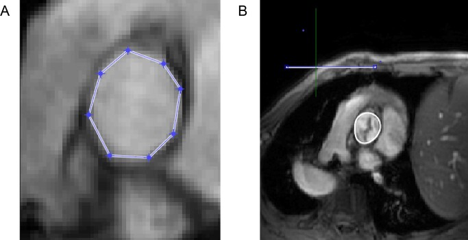
Magnetic resonance imaging provided the anatomical and velocity data required for the blood flow simulation. Patient-specific in- and outflow boundary conditions were used for the computational fluid dynamics analysis. Haemodynamic variables pertaining to blood flow pattern and derived from the magnetic resonance imaging data were calculated. However, we encountered problems in our multi-step methodology, most notably processing the flow data. This meant that other variables requiring computation with computational fluid dynamics could not be calculated.
Creating a model for computational fluid dynamics analysis is as complex as the physiology under scrutiny. We discuss some of the difficulties associated with creating such models, along with suggestions for improvements in order to yield reliable and beneficial results.
现代成像技术为计算流体动力学提供了越来越详细的解剖和生理信息,以预测生理现象的行为。计算机建模有助于规划合适的干预措施。我们小组使用磁共振成像和计算流体动力学来研究单纯组织主动脉瓣置换术前和术后二叶式主动脉瓣患者升主动脉中的血流动力学变量。计算机建模需要将生理模型转化为数学模型,通过在四个维度上进行多次迭代的方程来求解。创建这些模型涉及多个步骤且需要人工输入,这使得该过程容易出错,并限制了其在不同操作员之间以及同一操作员内部的可重复性。尽管存在这些挑战,我们还是为每位患者创建了计算模型,以研究手术前后升主动脉的血流情况。
磁共振成像提供了血流模拟所需的解剖和速度数据。针对每位患者的流入和流出边界条件用于计算流体动力学分析。计算了与血流模式相关且源自磁共振成像数据的血流动力学变量。然而,我们在多步骤方法中遇到了问题,最明显的是处理流量数据。这意味着其他需要用计算流体动力学进行计算的变量无法算出。
创建用于计算流体动力学分析的模型与所研究的生理过程一样复杂。我们讨论了与创建此类模型相关的一些困难,以及为获得可靠且有益的结果而提出的改进建议。