Hara Daisuke, Kondo Ryoichi, Shomura Toshitaka, Agatsuma Toshihiko, Saito Gaku
Department of Thoracic Surgery, National Hospital Organization Shinshu Ueda Medical Center, Nagano, Japan.
Division of General Thoracic Surgery, Department of Surgery, Shinshu University School of Medicine, Nagano, Japan.
Respir Med Case Rep. 2023 Jan 25;42:101817. doi: 10.1016/j.rmcr.2023.101817. eCollection 2023.
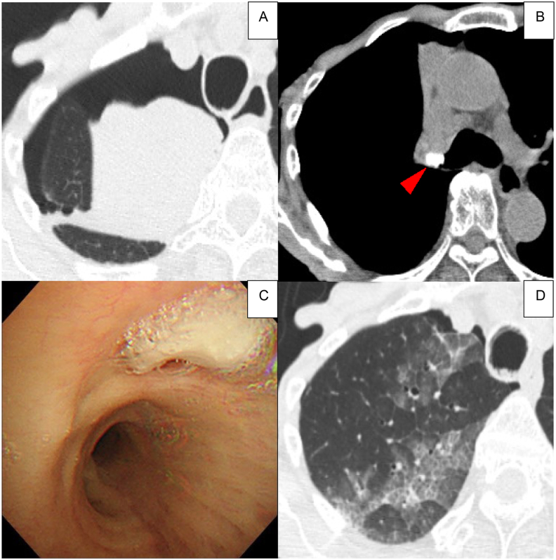
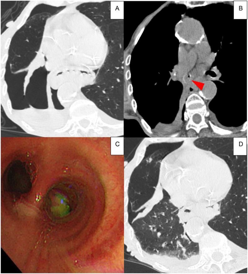
We report two cases of pulmonary collapse that simulated pneumothorax on computed tomographic images and were caused by rapid complete bronchial obstruction. One patient was a 77-year-old woman with sudden dyspnea, and the other was an 83-year-old woman with sudden dyspnea who was infected with influenza A virus. Chest computed tomography revealed lobular complete atelectasis with an almost complete expansion of the other lobes of the right lung. Some air space in the right pleural cavity was also observed. Both cases were diagnosed as "pneumothorax" by primary doctors. We noted the disappearance of air density in the lumen of the right bronchus in both cases. We performed bronchoscopy before thoracic drainage and removed the obstruction. Immediately, the obstructed pulmonary lobes expanded, and the air space in the pleural cavity disappeared without thoracic drainage. In the literature, this pneumothorax-like pulmonary collapse is called as "pneumothorax ex vacuo."
我们报告了两例在计算机断层扫描图像上模拟气胸的肺萎陷病例,其由快速完全性支气管阻塞引起。一例患者为一名77岁突发呼吸困难的女性,另一例为一名83岁感染甲型流感病毒且突发呼吸困难的女性。胸部计算机断层扫描显示小叶性完全肺不张,右肺其他叶几乎完全膨胀。右侧胸腔内也观察到一些气腔。两名患者最初均被主治医生诊断为“气胸”。我们注意到两例患者右支气管腔内空气密度均消失。我们在胸腔引流前进行了支气管镜检查并清除了阻塞物。随即,阻塞的肺叶扩张,胸腔内气腔未行胸腔引流即消失。在文献中,这种类似气胸的肺萎陷被称为“肺萎陷性气胸”。