Dwiyanti Stephani
Department of Dental Medicine, School of Medicine and Health Sciences, Atma Jaya Catholic University of Indonesia, Jakarta, Indonesia.
Case Rep Dent. 2023 Jan 23;2023:3024231. doi: 10.1155/2023/3024231. eCollection 2023.
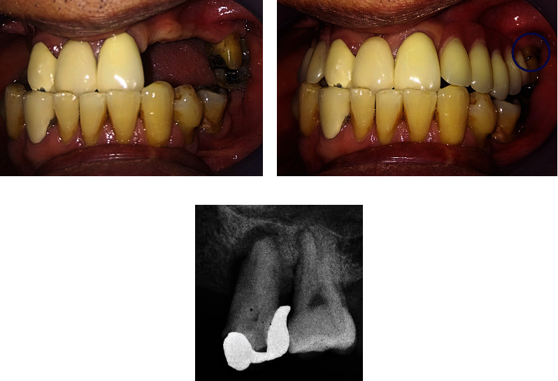
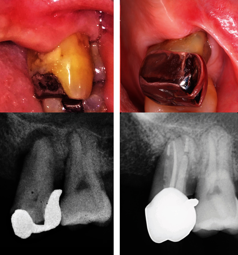
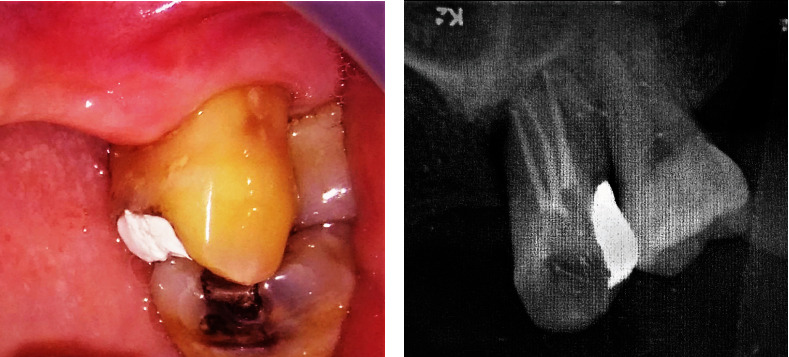
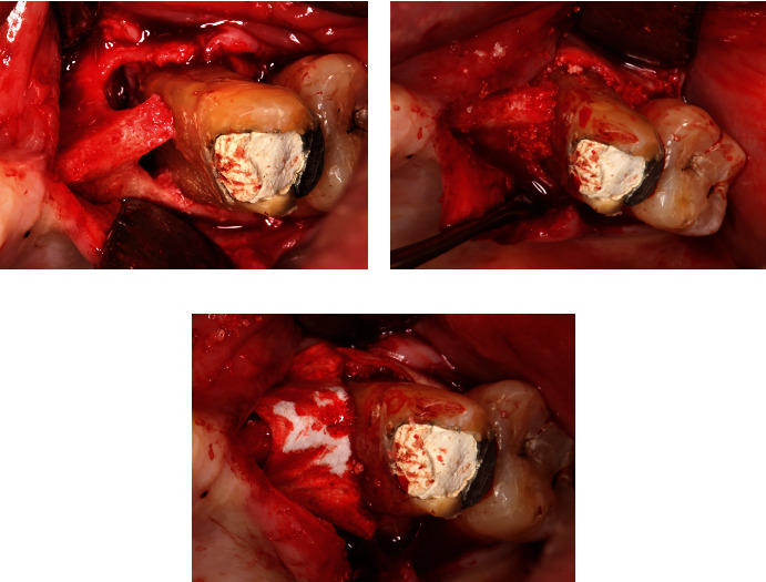
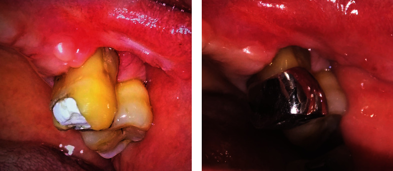
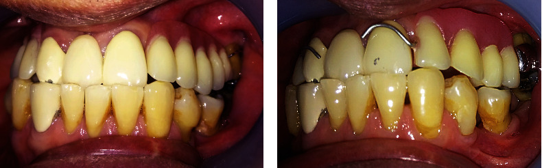
. This case report discusses the multi-disciplinary approach and long-term follow-up of a 66-year-old male who suffered a combined endodontic-periodontal lesion (EPL). As EPL is uncommon in daily practice and dentists' knowledge and awareness of EPL is quite low, this case becomes of high interest and value to document and research. The objective of this study is to present the diagnosis, multi-disciplinary approach, and long-term follow-up of compromised teeth with EPL. It highlights the importance of the identification and elimination of all causative factors as well as the correct treatment sequence to achieve a predictable outcome. The patient was referred to the periodontist after multiple unsuccessful attempts by his previous dentist. He complained of recurrent dull pain and abscess on his upper left tooth (tooth #26) that had been present for the past three years. A diagnosis of combined EPL was obtained after thorough anamnesis, clinical evaluation, and radiographic examination. The clinician identified several predisposing factors, such as plaque, trauma from occlusion, and excessive force on tooth #26 due to incorrect denture design. Treatment involved multiple dental specialties. At the periodontist, the patient underwent scaling, root planning, and removal of overhanging part of the restoration. At the endodontist, root canal treatment (RCT) was completed. Two months after RCT, a periodontal regenerative procedure was done. The defect was filled with a combination of allograft/alloplastic bone graft and covered with a barrier membrane. Upon healing, the prosthodontist did the final restoration of fiber post with a metal crown on tooth #26 and constructed a new denture with a periodontal-friendly design. A follow-up at five months and four years showed excellent results. The patient was symptom-free, and tooth #26 showed no periodontal inflammation. Radiographic examination showed a good bone fill at the defect. Supportive periodontal therapy should be emphasized to achieve the long-term success of EPL.
本病例报告探讨了一名66岁男性患牙髓-牙周联合病变(EPL)后的多学科治疗方法及长期随访情况。由于EPL在日常临床实践中并不常见,且牙医对其认识和了解程度较低,因此该病例对于记录和研究具有很高的价值。本研究的目的是介绍患有EPL的患牙的诊断、多学科治疗方法及长期随访情况。它强调了识别和消除所有致病因素以及正确的治疗顺序对于获得可预测结果的重要性。该患者在其前一位牙医多次治疗失败后被转诊至牙周病科医生处。他主诉左上牙(26号牙)反复出现钝痛和脓肿,这种情况已持续三年。经过全面的病史采集、临床评估和影像学检查后,确诊为牙髓-牙周联合病变。临床医生识别出了几个易感因素,如菌斑、咬合创伤以及由于义齿设计不当导致的26号牙受力过大。治疗涉及多个牙科专业领域。在牙周病科,患者接受了龈上洁治、根面平整以及去除修复体悬突部分的治疗。在牙髓病科,完成了根管治疗(RCT)。根管治疗两个月后,进行了牙周再生手术。缺损处用同种异体骨/人工骨移植材料联合填充,并覆盖屏障膜。愈合后,修复科医生对26号牙进行了纤维桩加金属冠的最终修复,并制作了一副牙周友好型设计的新义齿。五个月和四年后的随访结果显示效果极佳。患者无症状,26号牙无牙周炎症。影像学检查显示缺损处骨填充良好。应强调支持性牙周治疗以实现牙髓-牙周联合病变的长期成功治疗。