Müller Kilian, Berking Carola, Voskens Caroline, Heppt Markus V, Heinzerling Lucie, Koch Elias A T, Kramer Rafaela, Merkel Susanne, Schuler-Thurner Beatrice, Schellerer Vera, Steeb Theresa, Wessely Anja, Erdmann Michael
Institute of Hygiene and Environmental Medicine, University Medicine Greifswald, Greifswald, Germany.
Department of Dermatology, Uniklinikum Erlangen, Deutsches Zentrum Immuntherapie (DZI), Friedrich-Alexander University Erlangen-Nürnberg (FAU), Erlangen, Germany.
Front Med (Lausanne). 2023 Jan 19;10:1089013. doi: 10.3389/fmed.2023.1089013. eCollection 2023.
In melanoma, in-transit metastases characteristically occur at the lower extremity along lymphatic vessels.
The objective of this study was to evaluate conventional or three-dimensional photography as a tool to analyze in-transit metastasis pattern of melanoma of the lower extremity. In addition, we assessed risk factors for the development of in-transit metastases in cutaneous melanoma.
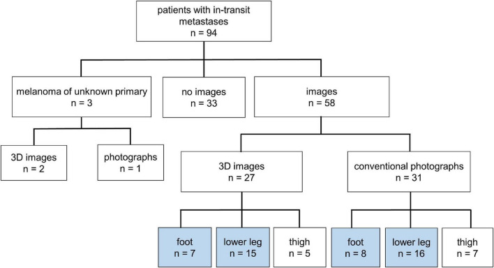
In this retrospective, monocentric study first we compared the clinical data of all evaluable patients with in-transit metastases of melanoma on the lower extremity ( = 94) with melanoma patients without recurrence of disease ( = 288). In addition, based on conventional ( = 24) and three-dimensional photography ( = 22), we defined the specific distribution patterns of the in-transit metastases on the lower extremity.
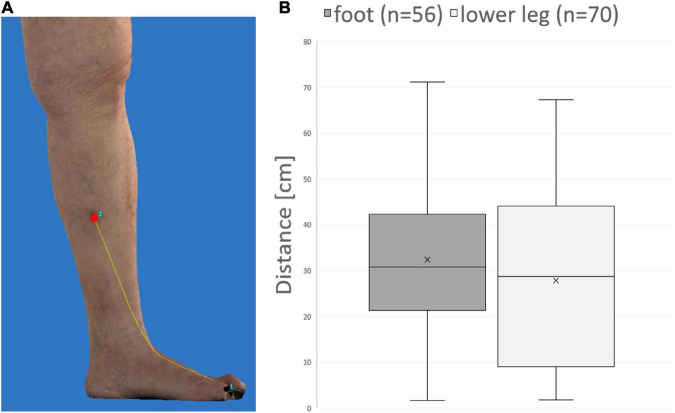
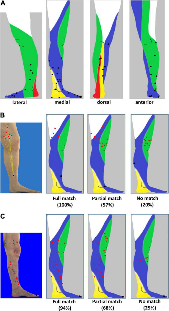
Using a multivariate analysis we identified nodular melanoma, tumor thickness, and ulceration as independent risk factors to develop in-transit metastases ITM ( = 94). In patients with melanoma on the lower leg ( = 31), in-transit metastases preferentially developed along anatomically predefined lymphatic pathways. In contrast when analyzing in-transit metastases of melanoma on the foot ( = 15) no clear pattern could be visualized. In addition, no difference in distance between in-transit metastases and primary melanoma on the foot compared to the lower leg was observed using three-dimensional photography ( = 22).
A risk-adapted follow-up of melanoma patients to detect in-transit metastases can be applied by knowledge of the specific lymphatic drainage of the lower extremity. Our current analysis suggests a more complex lymphatic drainage of the foot.
在黑色素瘤中,皮下转移瘤通常沿着淋巴管出现在下肢。
本研究的目的是评估传统摄影或三维摄影作为分析下肢黑色素瘤皮下转移模式的工具。此外,我们评估了皮肤黑色素瘤发生皮下转移的危险因素。
在这项回顾性单中心研究中,我们首先比较了所有可评估的下肢黑色素瘤皮下转移患者(n = 94)与无疾病复发的黑色素瘤患者(n = 288)的临床数据。此外,基于传统摄影(n = 24)和三维摄影(n = 22),我们定义了下肢皮下转移瘤的特定分布模式。
通过多变量分析,我们确定结节状黑色素瘤、肿瘤厚度和溃疡是发生皮下转移(ITM,n = 94)的独立危险因素。在小腿黑色素瘤患者(n = 31)中,皮下转移瘤优先沿着解剖学上预先定义的淋巴途径发展。相比之下,在分析足部黑色素瘤的皮下转移(n = 15)时,未观察到清晰的模式。此外,使用三维摄影(n = 22)观察到,足部皮下转移瘤与原发性黑色素瘤之间的距离与小腿相比没有差异。
通过了解下肢特定的淋巴引流情况,可以对黑色素瘤患者进行风险适应性随访以检测皮下转移瘤。我们目前的分析表明足部的淋巴引流更为复杂。