Sgouros Dimitrios, Theofili Melpomeni, Zafeiropoulou Theodora, Lallas Aimilios, Apalla Zoe, Zaras Alexios, Liopyris Konstantinos, Pappa Georgia, Polychronaki Eleni, Kousta Fiori, Panagiotopoulos Antonios, Stratigos Alexander, Rigopoulos Dimitrios, Katoulis Alexander C
2nd Department of Dermatology and Venereology, "Attikon" General University Hospital, Medical School, National and Kapodistrian University of Athens, 12462 Athens, Greece.
1st Department of Dermatology and Venereology, "Andreas Sygros" Hospital, Medical School, National and Kapodistrian University of Athens, 16121 Athens, Greece.
J Clin Med. 2023 Jan 30;12(3):1063. doi: 10.3390/jcm12031063.
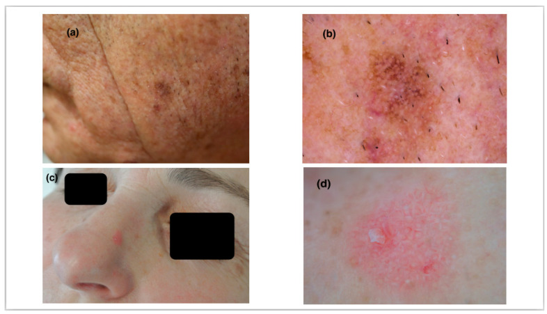
Dermoscopic features of actinic keratosis (AK) have been widely studied, but there is still little evidence for their diagnostic accuracy. Our study investigates whether established dermoscopic criteria are reliable predictors in differentiating non-pigmented actinic keratosis (NPAK) from pigmented actinic keratosis (PAK). For this purpose, dermoscopic images of 83 clinically diagnosed AK (45 NPAK, 38PAK) were examined, and the sensitivity (Se), specificity (Sp), positive predictive value (PPV), and negative predictive value (NPV) were assessed. Features with statistical significance were the red pseudo-network ( = 0.02) for NPAK and the pigmented pseudo-network ( < 0.001) with a pigment intensity value even less than 10% for PAK ( = 0.001). Pigmented pseudo-network (Se: 89%, Sp: 77%, PPV: 77%, NPV: 89%) with a pigment intensity value of more than 10% (Se: 90%, Sp: 86%, PPV: 79%, NPV: 93%) had excellent diagnostic accuracy for PAK. Scale and widened follicular openings with yellowish dots surrounded by white circles were equally represented in both variants of AK. Linear wavy vessels and shiny streaks were more prominently observed in NPAK, as were rosettes in PAK, but these results failed to meet statistical significance. The red starburst pattern was near statistical significance for PAK. Therefore, pigmentation is the strongest dermoscopic predictor for the differentiation between NPAK and PAK.
光化性角化病(AK)的皮肤镜特征已得到广泛研究,但关于其诊断准确性的证据仍然很少。我们的研究调查了既定的皮肤镜标准在区分非色素性光化性角化病(NPAK)和色素性光化性角化病(PAK)方面是否为可靠的预测指标。为此,检查了83例临床诊断为AK的皮肤镜图像(45例NPAK,38例PAK),并评估了敏感性(Se)、特异性(Sp)、阳性预测值(PPV)和阴性预测值(NPV)。具有统计学意义的特征是NPAK的红色假网络(P = 0.02)以及PAK的色素性假网络(P < 0.001),其色素强度值甚至低于10%(P = 0.001)。色素强度值超过10%的色素性假网络(Se:89%,Sp:77%,PPV:77%,NPV:89%)对PAK具有出色的诊断准确性。鳞屑和毛囊开口增宽伴白色圆圈环绕的淡黄色小点在AK的两种变体中均有同等表现。线性波浪状血管和发亮条纹在NPAK中更显著,PAK中的玫瑰花结也是如此,但这些结果未达到统计学意义。红色星芒状模式对PAK接近统计学意义。因此,色素沉着是区分NPAK和PAK的最强皮肤镜预测指标。