Department of Orthodontics, Shanghai Ninth People's Hospital, Shanghai Jiao Tong University School of Medicine;College of Stomatology, Shanghai Jiao Tong University; National Center for Stomatology; National Clinical Research Center for Oral Diseases; Shanghai Key Laboratory of Stomatology; Shanghai Research Institute of Stomatology, 500 Quxi Road, Shanghai, 200011, China.
Translational Medicine Research Platform of Oral Biomechanics and Artificial Intelligence, Department of Orthodontics, Shanghai Ninth People's Hospital, Shanghai Jiao Tong University School of Medicine;College of Stomatology, Shanghai Jiao Tong University; National Center for Stomatology; National Clinical Research Center for Oral Diseases; Shanghai Key Laboratory of Stomatology; Shanghai Research Institute of Stomatology, Shanghai, 200011, China.
Prog Orthod. 2023 Feb 13;24(1):5. doi: 10.1186/s40510-023-00454-7.
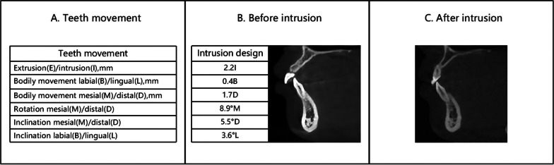
During the intrusion of lower incisors with clear aligners (CAs), root disengagement from the alveolar bone often occurs, resulting in serious complications. This study aimed to determine the potential force mechanism of the mandibular anterior teeth under the pressure of CA, providing theoretical data for clinical practice.
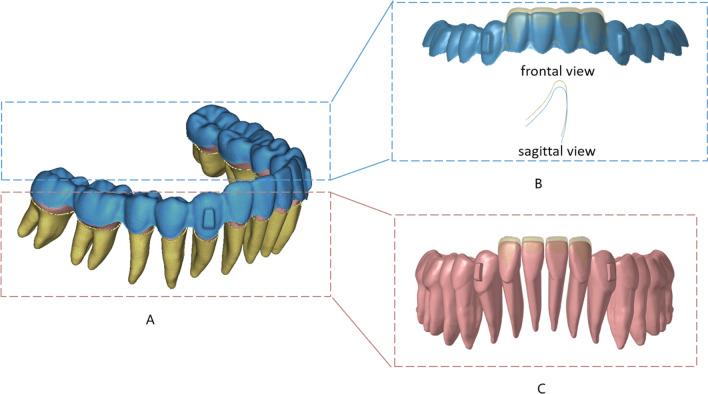
In this study, a 3D finite element model was established, including the CA, periodontal ligament, and mandibular dentition. Incisor mandibular plane angles were set as 5 groups: 90°, 95°, 100°, 105°, and 110°. The 4 mandibular incisors were intruded by 0.2 mm, while the canines were the anchorage teeth. The stress, force systems, and potential movement trends of mandibular anterior teeth were obtained.
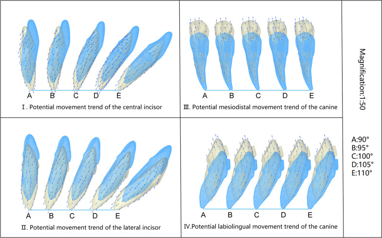
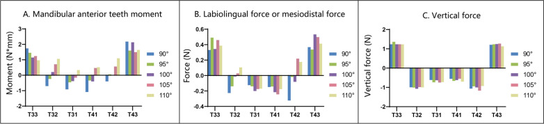
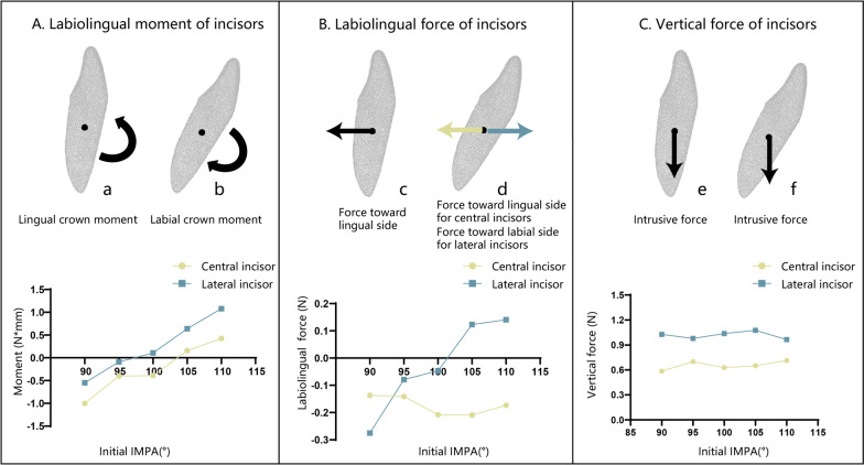
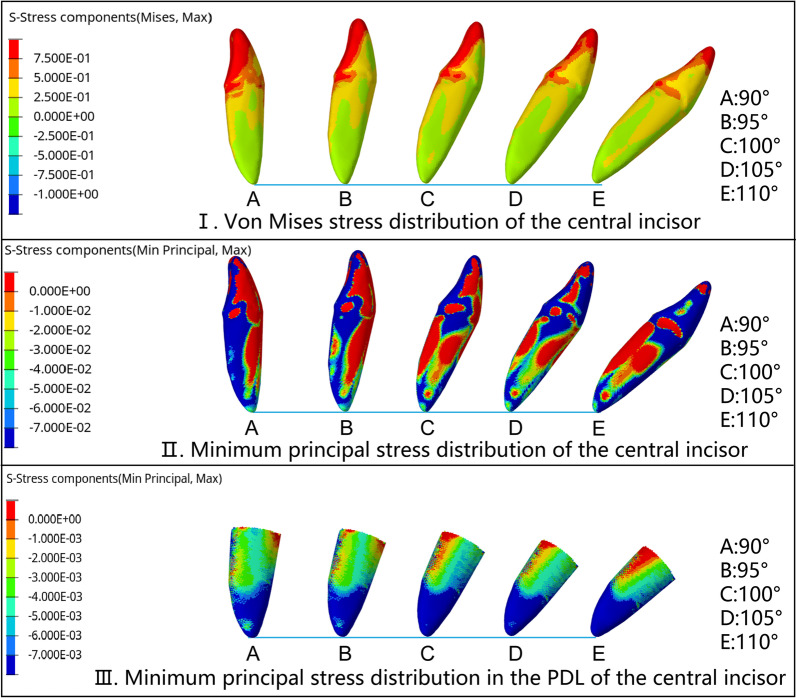
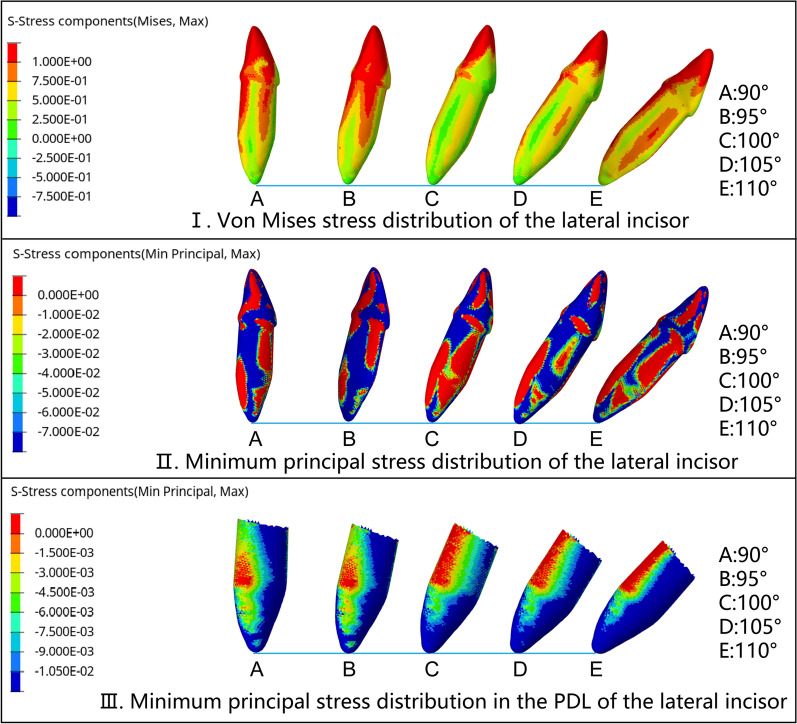
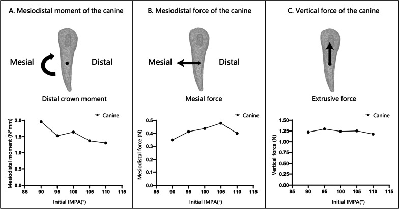
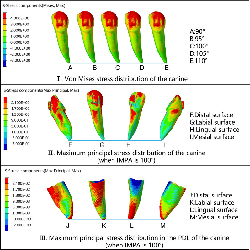
The compressive stress of the incisors was concentrated in the lingual fossa, incisal ridge, and apex. With the increase in IMPA, the moment of central incisors changed from lingual crown moment to labial crown moment, with the turning point between 100° and 105°, but the center of resistance (CR) was always subjected to the force toward the lingual and intrusive direction. The force and moment toward the labial side of the lateral incisors were greater than those toward the central incisors. The canines always tipped distally and received extrusive force with no relationship with IMPA.
With the increase in the initial IMPA, the direction of labiolingual force on the mandibular incisors was reversed. However, the root of the lower incisors always tipped labially, which indicated fenestration and dehiscence.
在使用透明矫正器(CA)内收下颌切牙时,牙根常常会从牙槽骨中脱离,导致严重的并发症。本研究旨在确定 CA 对下颌前牙施加压力时的潜在力机制,为临床实践提供理论数据。
本研究建立了一个包括 CA、牙周韧带和下颌牙列的三维有限元模型。切牙下颌平面角分为 5 组:90°、95°、100°、105°和 110°。用 0.2mm 的力内收 4 个下颌切牙,尖牙作为支抗牙。获得了下颌前牙的应力、力系统和潜在运动趋势。
切牙的压应力集中在舌窝、切缘和根尖。随着 IMPA 的增加,中切牙的力矩从舌侧冠力矩变为唇侧冠力矩,转折点在 100°到 105°之间,但中心阻力(CR)始终受到向舌侧和内收方向的力。侧切牙向唇侧的力和力矩大于中切牙。尖牙总是向远中倾斜并受到外展力,与 IMPA 无关。
随着初始 IMPA 的增加,下颌切牙的唇舌向力的方向发生反转。然而,下颌切牙的牙根总是向唇侧倾斜,这表明有开窗和骨开裂的风险。