Villalba Nuria, Ma Yonggang, Gahan Sarah A, Joly-Amado Aurelie, Spence Sam, Yang Xiaoyuan, Nash Kevin, Yuan Sarah Y
University of South Florida Morsani College of Medicine.
Res Sq. 2023 Jan 30:rs.3.rs-2511441. doi: 10.21203/rs.3.rs-2511441/v1.
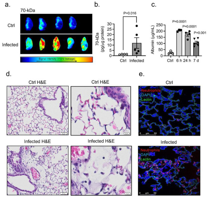
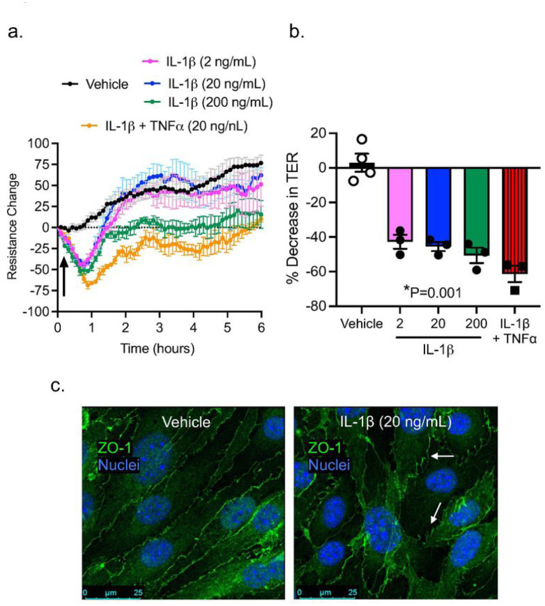
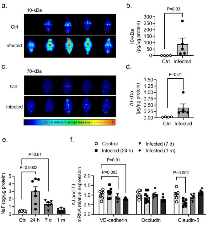
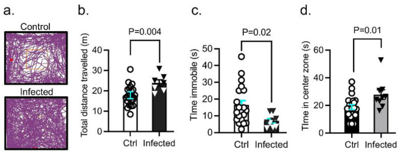
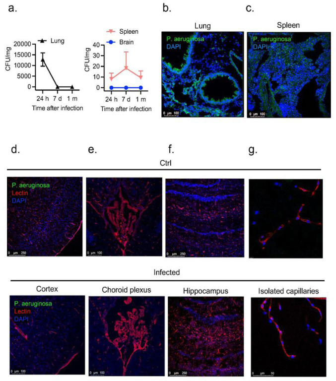
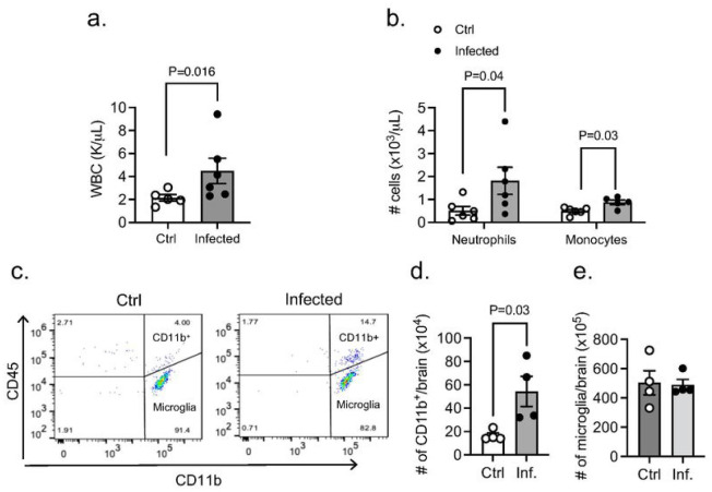
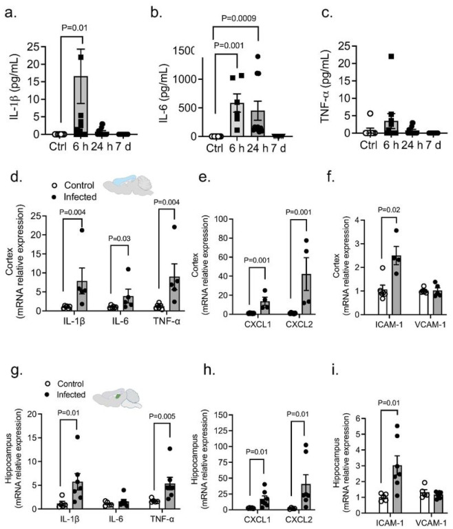
Background Severe lung infection can lead to brain dysfunction and neurobehavioral disorders. The mechanisms that regulate the lung-brain axis of inflammatory response to respiratory infection are incompletely understood. This study examined the effects of lung infection causing systemic and neuroinflammation as a potential mechanism contributing to blood-brain barrier (BBB) leakage and behavioral impairment. Methods Pneumonia was induced in adult C57BL/6 mice by intratracheal inoculation of (PA). Solute extravasation, histology, immunofluorescence, RT-PCR, multiphoton imaging and neurological testing were performed in this study. Results Lung infection caused alveolar-capillary barrier injury as indicated by leakage of plasma proteins across pulmonary microvessels and histopathological characteristics of pulmonary edema (alveolar wall thickening, microvessel congestion, and neutrophil infiltration). PA also caused significant BBB dysfunction characterized by leakage of different sized molecules across cerebral microvessels and a decreased expression of cell-cell junctions (VE-cadherin, claudin-5) in the brain. BBB leakage peaked at 24 hours and lasted for 7 days post-inoculation. Additionally, mice with lung infection displayed hyperlocomotion and anxiety-like behaviors. To test whether cerebral dysfunction was caused by PA directly or indirectly, we measured bacterial load in multiple organs. While PA loads were detected in the lungs up to 7 days post-inoculation, bacteria were not detected in the brain as evidenced by negative cerebral spinal fluid (CSF) cultures and lack of distribution in different brain regions or isolated cerebral microvessels. However, mice with PA lung infection demonstrated increased mRNA expression in the brain of pro-inflammatory cytokines (IL-1β, IL-6, and TNF-α), chemokines (CXCL-1, CXCL-2) and adhesion molecules (VCAM-1 and ICAM-1) along with CD11b + cell recruitment, corresponding to their increased blood levels of white cells (polymorphonuclear cells) and cytokines. To confirm the direct effect of cytokines on endothelial permeability, we measured cell-cell adhesive barrier resistance and junction morphology in mouse brain microvascular endothelial cell monolayers, where administration of IL-1β induced a significant reduction of barrier function coupled with tight junction (TJ) diffusion and disorganization. Combined treatment with IL-1β and TNFα augmented the barrier injury. Conclusions These results suggest that lung bacterial infection causes cerebral microvascular leakage and neuroinflammation via a mechanism involving cytokine-induced BBB injury.
背景 严重肺部感染可导致脑功能障碍和神经行为紊乱。对呼吸道感染炎症反应的肺-脑轴调节机制尚未完全了解。本研究探讨肺部感染引起全身和神经炎症作为导致血脑屏障(BBB)渗漏和行为障碍的潜在机制的作用。方法 通过气管内接种铜绿假单胞菌(PA)诱导成年C57BL/6小鼠发生肺炎。本研究进行了溶质外渗、组织学、免疫荧光、RT-PCR、多光子成像和神经学测试。结果 肺部感染导致肺泡-毛细血管屏障损伤,表现为血浆蛋白跨肺微血管渗漏以及肺水肿的组织病理学特征(肺泡壁增厚、微血管充血和中性粒细胞浸润)。PA还导致显著的血脑屏障功能障碍,表现为不同大小分子跨脑微血管渗漏以及脑中细胞间连接(血管内皮钙黏蛋白、闭合蛋白-5)表达降低。血脑屏障渗漏在接种后24小时达到峰值,并持续7天。此外,肺部感染的小鼠表现出运动亢进和焦虑样行为。为了测试脑功能障碍是由PA直接还是间接引起的,我们测量了多个器官中的细菌载量。虽然接种后7天内在肺部检测到PA载量,但脑脊髓液(CSF)培养阴性以及在不同脑区或分离的脑微血管中未检测到细菌,证明脑中未检测到细菌。然而,PA肺部感染的小鼠脑内促炎细胞因子(IL-1β、IL-6和TNF-α)、趋化因子(CXCL-1、CXCL-2)和黏附分子(VCAM-1和ICAM-1)的mRNA表达增加,同时伴有CD11b +细胞募集,这与它们血液中白细胞(多形核细胞)和细胞因子水平升高相对应。为了证实细胞因子对内皮通透性的直接作用,我们测量了小鼠脑微血管内皮细胞单层中的细胞间黏附屏障电阻和连接形态,其中给予IL-1β导致屏障功能显著降低,同时紧密连接(TJ)扩散和紊乱。IL-1β和TNFα联合治疗加剧了屏障损伤。结论 这些结果表明,肺部细菌感染通过细胞因子诱导的血脑屏障损伤机制导致脑微血管渗漏和神经炎症。