Zhang Xun, Hua Zhaohui, Chen Rui, Jiao Zhouyang, Shan Jintao, Li Chong, Li Zhen
Department of Endovascular Surgery, First Affiliated Hospital of Zhengzhou University, Zhengzhou, Henan, China.
Department of Magnetic Resonance Imaging, First Affiliated Hospital of Zhengzhou University, Zhengzhou, Henan, China.
Front Neurol. 2023 Jan 26;14:1050899. doi: 10.3389/fneur.2023.1050899. eCollection 2023.
Identification of vulnerable carotid plaque is important for the treatment and prevention of stroke. In previous studies, plaque vulnerability was assessed qualitatively. We aimed to develop a 3D carotid plaque radiomics model based on high-resolution magnetic resonance imaging (HRMRI) to quantitatively identify vulnerable plaques.
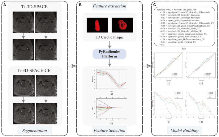
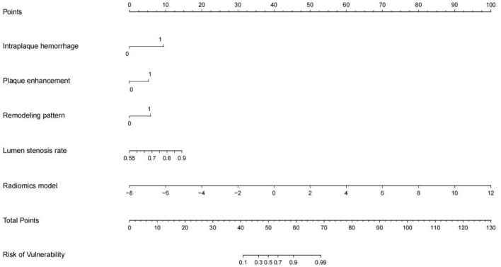
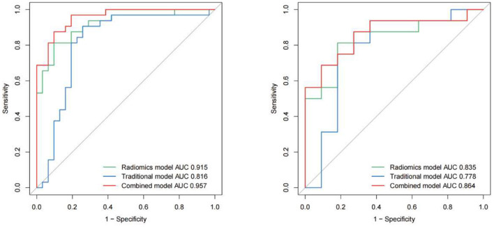
Ninety patients with carotid atherosclerosis who underwent HRMRI were randomized into training and test cohorts. Using the radiological characteristics of carotid plaques, a traditional model was constructed. A 3D carotid plaque radiomics model was constructed using the radiomics features of 3D T-SPACE and its contrast-enhanced sequences. A combined model was constructed using radiological and radiomics characteristics. Nomogram was generated based on the combined models, and ROC curves were utilized to assess the performance of each model.
48 patients (53.33%) were symptomatic and 42 (46.67%) were asymptomatic. The traditional model was constructed using intraplaque hemorrhage, plaque enhancement, wall remodeling pattern, and lumen stenosis, and it provided an area under the curve (AUC) of 0.816 vs. 0.778 in the training and testing sets. In the two cohorts, the 3D carotid plaque radiomics model and the combined model had an AUC of 0.915 vs. 0.835 and 0.957 vs. 0.864, respectively. In the training set, both the radiomics model and the combination model outperformed the traditional model, but there was no significant difference between the radiomics model and the combined model.
HRMRI-based 3D carotid radiomics models can improve the precision of detecting vulnerable carotid plaques, consequently improving risk classification and clinical decision-making in patients with carotid stenosis.
识别易损性颈动脉斑块对于中风的治疗和预防至关重要。在以往的研究中,斑块易损性是通过定性评估的。我们旨在基于高分辨率磁共振成像(HRMRI)开发一种三维颈动脉斑块影像组学模型,以定量识别易损斑块。
90例接受HRMRI检查的颈动脉粥样硬化患者被随机分为训练组和测试组。利用颈动脉斑块的放射学特征构建传统模型。利用三维T-SPACE及其对比增强序列的影像组学特征构建三维颈动脉斑块影像组学模型。利用放射学和影像组学特征构建联合模型。基于联合模型生成列线图,并利用ROC曲线评估各模型的性能。
48例(53.33%)有症状,42例(46.67%)无症状。传统模型利用斑块内出血、斑块强化、管壁重塑模式和管腔狭窄构建,在训练集和测试集中的曲线下面积(AUC)分别为0.816和0.778。在两个队列中,三维颈动脉斑块影像组学模型和联合模型的AUC分别为0.915对0.835和0.957对0.864。在训练集中,影像组学模型和联合模型均优于传统模型,但影像组学模型和联合模型之间无显著差异。
基于HRMRI的三维颈动脉影像组学模型可提高检测易损性颈动脉斑块的精度,从而改善颈动脉狭窄患者的风险分类和临床决策。