• 文献检索
  • 文档翻译
  • 深度研究
  • 学术资讯
  • Suppr Zotero 插件Zotero 插件
  • 邀请有礼
  • 套餐&价格
  • 历史记录
应用&插件
Suppr Zotero 插件Zotero 插件浏览器插件Mac 客户端Windows 客户端微信小程序
定价
高级版会员购买积分包购买API积分包
服务
文献检索文档翻译深度研究API 文档MCP 服务
关于我们
关于 Suppr公司介绍联系我们用户协议隐私条款
关注我们

Suppr 超能文献

核心技术专利:CN118964589B侵权必究
粤ICP备2023148730 号-1Suppr @ 2026

文献检索

告别复杂PubMed语法,用中文像聊天一样搜索,搜遍4000万医学文献。AI智能推荐,让科研检索更轻松。

立即免费搜索

文件翻译

保留排版,准确专业,支持PDF/Word/PPT等文件格式,支持 12+语言互译。

免费翻译文档

深度研究

AI帮你快速写综述,25分钟生成高质量综述,智能提取关键信息,辅助科研写作。

立即免费体验

单回波与多回波磁敏感加权成像在检测脑动静脉分流中的比较:一项初步研究

Comparison of Single- and Multi-Echo Susceptibility-Weighted Imaging in Detecting Cerebral Arteriovenous Shunts: A Preliminary Study.

作者信息

Han Seung Wan, Shin Jae Ho, Ihn Yon Kwon, Yang Seung Ho, Sung Jae Hoon

出版信息

J Korean Soc Radiol. 2023 Jan;84(1):226-239. doi: 10.3348/jksr.2022.0080. Epub 2023 Jan 30.

DOI:10.3348/jksr.2022.0080
PMID:36818708
原文链接:https://pmc.ncbi.nlm.nih.gov/articles/PMC9935958/
Abstract

PURPOSE

To compare the sensitivities of T2-weighted image (T2WI) and susceptibility-weighted imaging (SWI) in detecting cerebral arteriovenous fistula (AVF), cerebral arteriovenous malformation (AVM), and carotid-cavernous sinus fistula (CCF), and to qualitatively evaluate single-echo SWI (s-SWI) and multi-echo SWI (m-SWI) in characterizing vascular lesions.

MATERIALS AND METHODS

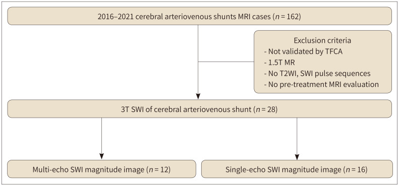
From January 2016 to December 2021, cerebral angiography-proven lesions were recruited. The sensitivities of T2WI and SWI in detecting vascular lesions were compared using McNemar's test. Qualitative evaluations of s-SWI and m-SWI were categorized to be of poor, average, or good quality and compared using Fisher's exact test.

RESULTS

A total of 24 patients (mean age: 61 years, 12 female, and 12 male) were enrolled. Twenty patients underwent s-SWI or m-SWI, and four patients underwent both. AVF, AVM, and CCF were diagnosed in 10, 11, and 3 patients, respectively. SWI demonstrated higher sensitivity compared to that of T2WI (82.1% vs. 53.6%, = 0.013). m-SWI showed better image quality compared to that of s-SWI (good quality, 83.3% vs. 25.0%, = 0.009).

CONCLUSION

SWI demonstrated a higher sensitivity for detecting cerebral arteriovenous shunts compared to that of T2WI. m-SWI exhibited better image quality compared to that of s-SWI in characterizing vascular lesions.

摘要

目的

比较T2加权成像(T2WI)和磁敏感加权成像(SWI)在检测脑动静脉瘘(AVF)、脑动静脉畸形(AVM)和颈内动脉海绵窦瘘(CCF)方面的敏感性,并定性评估单回波SWI(s-SWI)和多回波SWI(m-SWI)对血管病变的特征性表现。

材料与方法

纳入2016年1月至2021年12月期间经脑血管造影证实的病变。采用McNemar检验比较T2WI和SWI检测血管病变的敏感性。将s-SWI和m-SWI的定性评估分为质量差、一般或良好,并采用Fisher精确检验进行比较。

结果

共纳入24例患者(平均年龄:61岁,女性12例,男性12例)。20例患者接受了s-SWI或m-SWI检查,4例患者同时接受了两种检查。分别在10例、11例和3例患者中诊断出AVF、AVM和CCF。与T2WI相比,SWI显示出更高的敏感性(82.1%对53.6%,P = 0.013)。与s-SWI相比,m-SWI显示出更好的图像质量(良好质量,83.3%对25.0%,P = 0.009)。

结论

与T2WI相比,SWI在检测脑动静脉分流方面具有更高的敏感性。在对血管病变的特征性表现方面,m-SWI比s-SWI表现出更好的图像质量。

https://cdn.ncbi.nlm.nih.gov/pmc/blobs/a244/9935958/eb24ff025f71/jksr-84-226-g005.jpg
https://cdn.ncbi.nlm.nih.gov/pmc/blobs/a244/9935958/a27b9c6d2692/jksr-84-226-g001.jpg
https://cdn.ncbi.nlm.nih.gov/pmc/blobs/a244/9935958/a417de67aacf/jksr-84-226-g002.jpg
https://cdn.ncbi.nlm.nih.gov/pmc/blobs/a244/9935958/9ff5d05c09ed/jksr-84-226-g003.jpg
https://cdn.ncbi.nlm.nih.gov/pmc/blobs/a244/9935958/e6ae6600654a/jksr-84-226-g004.jpg
https://cdn.ncbi.nlm.nih.gov/pmc/blobs/a244/9935958/eb24ff025f71/jksr-84-226-g005.jpg

相似文献

1
Comparison of Single- and Multi-Echo Susceptibility-Weighted Imaging in Detecting Cerebral Arteriovenous Shunts: A Preliminary Study.单回波与多回波磁敏感加权成像在检测脑动静脉分流中的比较:一项初步研究
J Korean Soc Radiol. 2023 Jan;84(1):226-239. doi: 10.3348/jksr.2022.0080. Epub 2023 Jan 30.
2
Accuracy of SWI sequences compared to T2*-weighted gradient echo sequences in the detection of cerebral cavernous malformations in the familial form.在家族性脑海绵状畸形检测中,与T2*加权梯度回波序列相比,SWI序列的准确性。
Neuroradiol J. 2016 Oct;29(5):326-35. doi: 10.1177/1971400916665376. Epub 2016 Aug 22.
3
Susceptibility-weighted imaging for the evaluation of patients with familial cerebral cavernous malformations: a comparison with t2-weighted fast spin-echo and gradient-echo sequences.磁敏感加权成像用于评估家族性脑海绵状血管畸形患者:与T2加权快速自旋回波序列和梯度回波序列的比较
AJNR Am J Neuroradiol. 2008 Jan;29(1):154-8. doi: 10.3174/ajnr.A0748. Epub 2007 Oct 18.
4
The value of susceptibility weighted magnetic resonance imaging in evaluation of patients with familial cerebral cavernous angioma.susceptibility加权磁共振成像在家族性脑海绵状血管瘤患者评估中的价值
Int J Clin Exp Med. 2014 Dec 15;7(12):5296-302. eCollection 2014.
5
Magnetic resonance susceptibility weighted imaging in neurosurgery: current applications and future perspectives.神经外科中的磁共振 susceptibility 加权成像:当前应用与未来展望。
J Neurosurg. 2015 Dec;123(6):1463-75. doi: 10.3171/2015.1.JNS142349. Epub 2015 Jul 24.
6
Susceptibility-weighted imaging in carotido-cavernous fistulas. A case control study.颈动脉海绵窦瘘的磁敏感加权成像。一项病例对照研究。
Interv Neuroradiol. 2013 Dec;19(4):438-44. doi: 10.1177/159101991301900406. Epub 2013 Dec 18.
7
Evaluation of Angioarchitectural Features of Unruptured Brain Arteriovenous Malformation by Susceptibility Weighted Imaging.利用磁敏感加权成像评估未破裂脑动静脉畸形的血管构筑特征
World Neurosurg. 2018 Aug;116:e1015-e1022. doi: 10.1016/j.wneu.2018.05.151. Epub 2018 May 30.
8
Noncavernous arteriovenous shunts mimicking carotid cavernous fistulae.酷似颈动脉海绵窦瘘的非海绵窦动静脉分流
Diagn Interv Radiol. 2016 Nov-Dec;22(6):555-559. doi: 10.5152/dir.2016.16073.
9
Evaluation of cerebral arteriovenous shunts: a comparison of parallel imaging time-of-flight magnetic resonance angiography (TOF-MRA) and compressed sensing TOF-MRA to digital subtraction angiography.脑动静脉分流的评估:并行成像时间飞跃磁共振血管造影(TOF-MRA)和压缩感知TOF-MRA与数字减影血管造影的比较。
Neuroradiology. 2021 Jun;63(6):879-887. doi: 10.1007/s00234-020-02581-y. Epub 2020 Oct 15.
10
Intracranial dural arteriovenous fistula with retrograde cortical venous drainage: use of susceptibility-weighted imaging in combination with dynamic susceptibility contrast imaging.颅内硬脑膜动静脉瘘伴逆行皮质静脉引流:磁敏感加权成像结合动态磁敏感对比成像的应用。
AJNR Am J Neuroradiol. 2010 Nov;31(10):1903-10. doi: 10.3174/ajnr.A2231. Epub 2010 Sep 2.

本文引用的文献

1
Risk Factors of Aggressive Clinical Presentation in Patients with Angiographically Aggressive Cranial Dural Arteriovenous Fistulas.血管造影显示为侵袭性的颅内硬脑膜动静脉瘘患者出现侵袭性临床表现的危险因素
J Clin Med. 2021 Dec 13;10(24):5835. doi: 10.3390/jcm10245835.
2
Brain arteriovenous malformations: A review of natural history, pathobiology, and interventions.脑动静脉畸形:自然史、病理生物学和干预措施的综述。
Neurology. 2020 Nov 17;95(20):917-927. doi: 10.1212/WNL.0000000000010968. Epub 2020 Oct 1.
3
Thin-Section MR Imaging for Carotid Cavernous Fistula.
颈动脉海绵窦瘘的薄层磁共振成像
AJNR Am J Neuroradiol. 2020 Sep;41(9):1599-1605. doi: 10.3174/ajnr.A6757. Epub 2020 Aug 20.
4
Susceptibility-Based Characterization of Cerebral Arteriovenous Malformations.基于易感性的脑动静脉畸形特征描述。
Invest Radiol. 2020 Nov;55(11):702-710. doi: 10.1097/RLI.0000000000000695.
5
Susceptibility-Weighted Angiography for the Follow-Up of Brain Arteriovenous Malformations Treated with Stereotactic Radiosurgery.立体定向放射外科治疗脑动静脉畸形的随访中应用磁敏感加权血管成像。
AJNR Am J Neuroradiol. 2019 May;40(5):792-797. doi: 10.3174/ajnr.A6053. Epub 2019 Apr 25.
6
Arterial-spin labeling MRI identifies residual cerebral arteriovenous malformation following stereotactic radiosurgery treatment.动脉自旋标记 MRI 识别立体定向放射外科治疗后的残余脑动静脉畸形。
J Neuroradiol. 2020 Feb;47(1):13-19. doi: 10.1016/j.neurad.2018.12.004. Epub 2019 Jan 15.
7
Arterial Spin-Labeling Improves Detection of Intracranial Dural Arteriovenous Fistulas with MRI.动脉自旋标记术提高 MRI 对颅内硬脑膜动静脉瘘的检出率。
AJNR Am J Neuroradiol. 2018 Apr;39(4):669-677. doi: 10.3174/ajnr.A5570. Epub 2018 Mar 15.
8
Epidemiology, genetics, pathophysiology, and prognostic classifications of cerebral arteriovenous malformations.脑动静脉畸形的流行病学、遗传学、病理生理学及预后分类
Handb Clin Neurol. 2017;143:5-13. doi: 10.1016/B978-0-444-63640-9.00001-1.
9
Intracranial Dural Arteriovenous Fistulae.颅内硬脑膜动静脉瘘
Stroke. 2017 May;48(5):1424-1431. doi: 10.1161/STROKEAHA.116.012784.
10
Intracranial Arteriovenous Shunting: Detection with Arterial Spin-Labeling and Susceptibility-Weighted Imaging Combined.颅内动静脉分流:动脉自旋标记与磁敏感加权成像联合检测
AJNR Am J Neuroradiol. 2017 Jan;38(1):71-76. doi: 10.3174/ajnr.A4961. Epub 2016 Oct 27.