Mangold Melanie Scarlett, Rüber Fabienne, Steinack Carolin, Gautschi Fiorenza, Wani Jasmin, Grimaldi Sascha, Franzen Daniel Peter
Department of Pulmonology, University Hospital Zurich, Raemistrasse 100, 8091 Zurich, Switzerland.
Department of Internal Medicine, Spital Uster, Brunnenstrasse 42, 8610 Uster, Switzerland.
J Clin Med. 2023 Feb 12;12(4):1474. doi: 10.3390/jcm12041474.
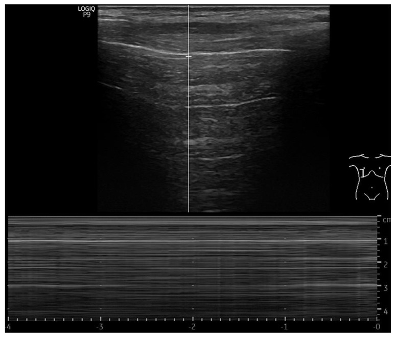
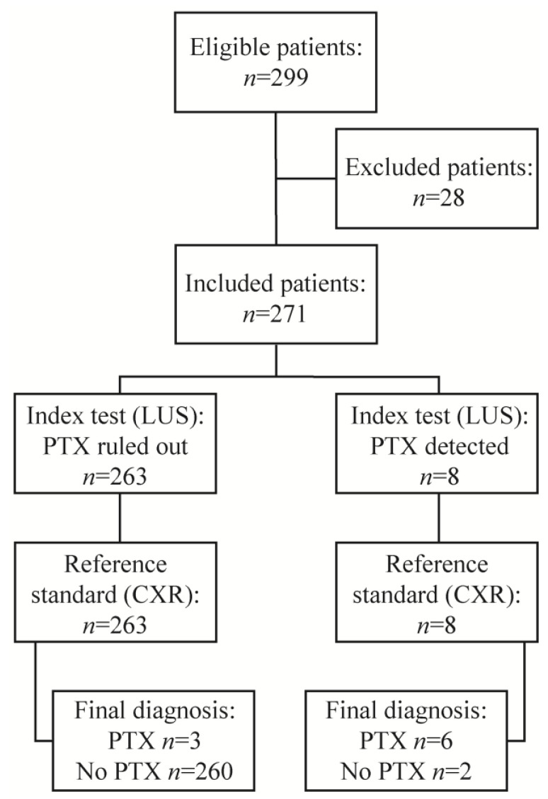
A chest X-ray (CXR) is recommended after bronchoscopies with an increased risk of pneumothorax (PTX). However, concerns regarding radiation exposure, expenses and staff requirements exist. A lung ultrasound (LUS) is a promising alternative for the detection of PTX, though data are scarce. This study aims to investigate the diagnostic yield of LUS compared to CXR, to exclude PTX after bronchoscopies with increased risk. This retrospective single-centre study included transbronchial forceps biopsies, transbronchial lung cryobiopsies and endobronchial valve treatments. Post-interventional PTX screening consisted of immediate LUS and CXR within two hours. In total, 271 patients were included. Early PTX incidence was 3.3%. Sensitivity, specificity, and the positive and negative predictive values of LUS were 67.7% (95% CI 29.93-92.51%), 99.2% (95% CI 97.27-99.91%), 75.0% (95% CI 41.16-92.79%) and 98.9% (95% CI 97.18-99.54%), respectively. PTX detection by LUS enabled the immediate placement of two pleural drains along with the bronchoscopy. With CXR, three false-positives and one false-negative were observed; the latter evolved into a tension-PTX. LUS correctly diagnosed these cases. Despite low sensitivity, LUS enables early diagnosis of PTX, thus preventing treatment delays. We recommend immediate LUS, in addition to LUS or CXR after two to four hours and monitoring for signs and symptoms. Prospective studies with higher sample sizes are needed.
对于气胸(PTX)风险增加的支气管镜检查,建议进行胸部X光(CXR)检查。然而,存在对辐射暴露、费用和人员需求的担忧。肺超声(LUS)是检测PTX的一种有前景的替代方法,尽管相关数据较少。本研究旨在调查LUS与CXR相比的诊断率,以排除气胸风险增加的支气管镜检查后的PTX。这项回顾性单中心研究包括经支气管钳夹活检、经支气管肺冷冻活检和支气管内瓣膜治疗。介入后PTX筛查包括在两小时内立即进行LUS和CXR。总共纳入了271例患者。早期PTX发生率为3.3%。LUS的敏感性、特异性、阳性和阴性预测值分别为67.7%(95%可信区间29.93 - 92.51%)、99.2%(95%可信区间97.27 - 99.91%)、75.0%(95%可信区间41.16 - 92.79%)和98.9%(95%可信区间97.18 - 99.54%)。通过LUS检测到PTX后,可在支气管镜检查的同时立即放置两根胸腔引流管。在CXR检查中,观察到3例假阳性和1例假阴性;后者发展为张力性PTX。LUS正确诊断了这些病例。尽管敏感性较低,但LUS能够早期诊断PTX,从而防止治疗延误。我们建议立即进行LUS检查,此外在两至四小时后进行LUS或CXR检查,并监测体征和症状。需要进行样本量更大的前瞻性研究。